 Indietro
Anatomy & Physiology: Cardiovascular and Blood System Fundamentals
 2:36

Termini in questo insieme (51)
Normal values of RBC
Functions of RBC
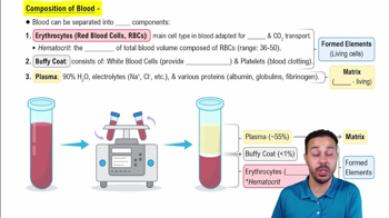
Normal values of RBC: 4.5-5.5 million/µL,
Functions of RBC: transport oxygen;
Normal Value of Platelets
Function of Platelets
Normal Value Platelets: 150,000-450,000/µL
Function of Platelets: blood clotting
Normal Values of Blood Volume
Adult Male: 5-6 Liters
Adult Female 4-5 Liters
Normal Values of Hematocrit
Functions of Hematocrit
Values of Hematocrit:
Adult Male 40-54% or 47% +/- 5%
Adult Female 38-46% or 42% +/- 5%
Function of Hematocrit: percentage of RBCs in blood;
Normal Values of WBC
Function of WBC
Value WBC: 4,000-11,000/µL,
Function of WBC: immune defense;
Normal Values of Hemoglobin
Function of Hemoglobin
Value of Hemoglobin: 12-16 g/dL (female), 14-18 g/dL (male).
Function of Hemoglobin: oxygen-carrying protein in RBCs.
Heart
blood vessels (arteries, veins, capillaries)
blood
Albumin is the most abundant plasma protein,
making up 60%,
helps maintain osmotic pressure.
Globin
Globin makes up 36% of plasma proteins;
Alpha and Beta globulins
produced by liver
functions as transport protein for metal ions ( Fe2+, Cu2+)
Gamma Globin in plasma proteins
produced by plasma cells
only protein not produced by the liver
acts as an antibody.
Include urea
creatinine
uric acid
ammonium salts
waste products filtered by kidneys.
Glucose
amino acids
fatty acids
vitamins
transported for cellular metabolism.
Leukopenia
low WBC count;
Leukocytosis
high WBC count, often due to infection.
Thrombus
blood clot attached to vessel wall;
Embolus
detached clot traveling in bloodstream.
Thrombocytopenia
low platelet count causing bleeding;
Hemophilia
genetic clotting factor deficiency.
Primary: platelet plug formation;
Secondary: coagulation cascade forming fibrin clot.
Platelets contract to shrink clot;
vessel repairs via endothelial cell growth.
Group A: A antigen, anti-B antibody;
Group B: B antigen, anti-A antibody;
Group AB: A & B antigens, no antibodies;
Group O: no antigens, anti-A & anti-B antibodies.
Donors must lack recipient antibodies;
O is universal donor,
AB universal recipient.
About the size of a fist,
located in the mediastinum
tilted left,
between lungs.
Fibrous pericardium protects;
serous pericardium has parietal and visceral layers with pericardial cavity reducing friction.
Epicardium: outer layer
Myocardium: muscular middle layer
Endocardium: inner lining.
Atrioventricular valves: tricuspid (right)
bicuspid/mitral (left)
Semilunar valves: pulmonary and aortic.
Superior/inferior vena cava to right atrium;
pulmonary trunk from right ventricle;
pulmonary veins to left atrium; aorta from left ventricle.
Left/right coronary arteries,
anterior/posterior interventricular arteries,
circumflex artery, coronary sinus, cardiac veins.
Ischemic: reduced blood flow;
angina: chest pain from ischemia;
infarction: tissue death from lack of blood.
Striated,
Involuntary,
branched cells with intercalated discs for synchronized contraction.
Autorhythmic cells generate impulses;
contractile cells perform contraction;
differ in action potential and ion permeability.