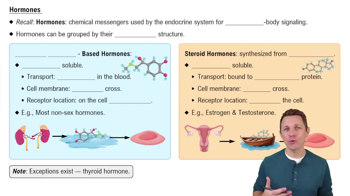
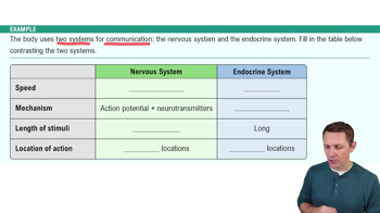
Primary, secondary, and secondary neuroendocrine organs
Primary organs produce hormones as their main function; secondary organs have other main functions but also secrete hormones; secondary neuroendocrine organs have neural and endocrine functions.
Anterior pituitary hormones
Includes hormones like GH, TSH, ACTH, FSH, LH, and PRL; they target various glands and tissues to regulate growth, metabolism, reproduction, and lactation.
Posterior pituitary hormones
Releases oxytocin and antidiuretic hormone (ADH), which regulate uterine contractions, milk ejection, and water balance.
Hypothalamus role in endocrine system
Controls the pituitary gland by producing releasing and inhibiting hormones; acts as a primary neuroendocrine organ.
Adrenal gland structure
Consists of a capsule, cortex (with three layers: zona glomerulosa, fasciculata, reticularis), and medulla producing steroid hormones and catecholamines.
Pancreas endocrine vs. exocrine portions
Islets of Langerhans are endocrine, secreting insulin and glucagon; acini are exocrine, producing digestive enzymes.
Type 1 vs. Type 2 diabetes
Type 1 is autoimmune destruction of beta cells causing insulin deficiency; Type 2 is insulin resistance with relative insulin deficiency.
Heart chambers and their function
Right atrium and ventricle pump deoxygenated blood to lungs; left atrium and ventricle pump oxygenated blood to the body.
Direction of blood flow through the heart
Blood flows: body → superior/inferior vena cava → right atrium → right ventricle → pulmonary trunk → lungs → pulmonary veins → left atrium → left ventricle → aorta → body.
Coronary arteries
Include the right coronary artery, left coronary artery, anterior interventricular artery, and circumflex artery; they supply oxygenated blood to the heart muscle.
Coronary veins
Include the great cardiac vein, middle cardiac vein, small cardiac vein, and coronary sinus; they drain deoxygenated blood from the heart muscle.
Cardiac muscle histology features
Contains intercalated discs for cell connection, myocytes as muscle cells, and centrally located myocyte nuclei.
ECG waves and their significance
P wave: atrial depolarization; QRS complex: ventricular depolarization; T wave: ventricular repolarization.
Cardiac output (CO) formula
CO = \(SV \times HR\), where SV is stroke volume and HR is heart rate.
Great vessels of the heart
Include the aorta, pulmonary trunk, pulmonary arteries, pulmonary veins, and superior and inferior vena cava.
Heart external anatomy landmarks
Includes the apex, base, left and right auricles, ligamentum arteriosum, and anterior interventricular sulcus.
Heart internal anatomy structures
Includes atria, ventricles, interventricular septum, pectinate muscles, trabeculae carneae, and valves.
Atrioventricular valves
The tricuspid valve on the right and the bicuspid (mitral) valve on the left prevent backflow into atria during ventricular contraction.
Chordae tendineae and papillary muscles
Chordae tendineae connect valve cusps to papillary muscles, preventing valve prolapse during ventricular contraction.
Heart tissue layers
From outer to inner: fibrous pericardium, serous pericardium (parietal and visceral layers), myocardium, and endocardium.
Cardiac tamponade
A condition where fluid accumulation in the pericardial cavity compresses the heart, impairing its function.
Fossa ovalis function
A remnant of the fetal foramen ovale, allowing blood to bypass the lungs before birth.
 Indietro
 7:06