 Indietro
Anatomy & Physiology: Endocrine and Cardiovascular Systems Study Guide
 7:06

Termini in questo insieme (30)
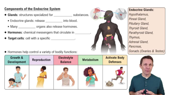
Ductless gland that secretes hormones directly into the bloodstream to act on distant target cells.
A specific cell possessing protein receptors that uniquely bind a particular hormone.
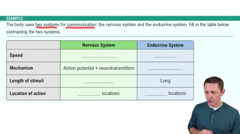
Chemical signal released into extracellular fluid to act on neighboring, different cell types.
Chemical signal that acts on the same cell or same cell type that secreted it.
Interaction where two or more hormones act together to produce an effect greater than the sum of their individual effects.
Interaction where one hormone opposes or inhibits the action of another hormone.
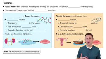
Hydrophilic hormones (amines, peptides, proteins) that travel freely in blood and bind to cell-surface receptors.
Lipophilic hormones derived from cholesterol that require transport proteins and bind to intracellular receptors.
Hydrophilic hormone binds membrane receptor → activates G-protein → activates adenylate cyclase → converts ATP to \(cAMP\) → activates Protein Kinase A to alter cell activity.
Lipophilic hormone diffuses into cell → binds intracellular receptor → hormone-receptor complex enters nucleus → alters gene transcription and protein synthesis.
Stalk connecting the hypothalamus to the pituitary gland.
Glandular tissue linked to hypothalamus via the Hypophyseal Portal System, secretes hormones like TSH, ACTH, LH, FSH, GH, and PRL.
Neural tissue storing and releasing hormones made in hypothalamus, including ADH and Oxytocin.
Produce \(T3\) and \(T4\), regulating basal metabolic rate, body temperature, and growth.
Secrete Calcitonin, which lowers blood calcium by inhibiting osteoclasts.
Produce Parathyroid Hormone (PTH), raising blood calcium by stimulating osteoclasts, increasing renal calcium reabsorption, and activating Vitamin D.
Zona Glomerulosa: Mineralocorticoids (Aldosterone); Zona Fasciculata: Glucocorticoids (Cortisol); Zona Reticularis: Androgens (DHEA).
Chromaffin cells secrete Epinephrine and Norepinephrine for sympathetic "fight-or-flight" responses.
Alpha cells produce Glucagon (raises blood glucose); Beta cells produce Insulin (lowers blood glucose).
Epicardium: visceral serous pericardium; Myocardium: cardiac muscle; Endocardium: inner endothelial lining.
Tricuspid valve on right; Bicuspid (Mitral) valve on left; Chordae tendineae prevent valve prolapse during ventricular contraction.
Pulmonary valve between right ventricle and pulmonary trunk; Aortic valve between left ventricle and aorta.
Phase 0: Na+ influx; Phase 1: initial K+ efflux; Phase 2: Ca2+ influx balances K+ efflux (plateau); Phase 3: K+ efflux; Phase 4: resting potential (~-90 mV).
Contain desmosomes for mechanical anchoring and gap junctions for electrical coupling of cardiomyocytes.
SA node → AV node (delay) → AV bundle → bundle branches → Purkinje fibers for coordinated contraction.
P wave: atrial depolarization; QRS complex: ventricular depolarization; T wave: ventricular repolarization; P-R interval: atrial to ventricular depolarization delay.
1. Ventricular filling (AV valves open); 2. Isovolumetric contraction (AV valves close, SL closed); 3. Ventricular ejection (SL valves open); 4. Isovolumetric relaxation (SL valves close).
SV = End-Diastolic Volume (EDV) − End-Systolic Volume (ESV)
CO = Heart Rate (HR) × Stroke Volume (SV)
Preload (sarcomere stretch), Contractility (strength of contraction), Afterload (arterial backpressure).