 Indietro
Anatomy & Physiology: Eye Structure and Visual Function
 4:16
4:16
Termini in questo insieme (24)
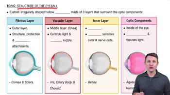
The outermost layer composed of dense avascular connective tissue, including the sclera and cornea. It protects and shapes the eyeball and anchors extrinsic eye muscles.
Opaque posterior region of the fibrous layer that protects the eyeball and anchors extrinsic eye muscles. Continuous with dura mater where the optic nerve exits.
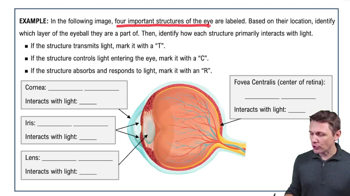
Transparent anterior one-sixth of the fibrous layer that allows light to enter and bends light. Covered by epithelium with pain receptors triggering blinking and tearing reflexes.
Middle pigmented layer consisting of the choroid, ciliary body, and iris. Supplies blood and absorbs light to prevent scattering.
Posterior portion of the uvea that supplies blood to all layers of the eyeball and contains brown pigment to absorb light.
Thickened ring of tissue surrounding the lens with ciliary muscles that control lens shape and ciliary processes that secrete aqueous humor.
The iris is the colored part of the eye controlling pupil size. The pupil regulates light entry: sphincter pupillae constricts it under parasympathetic control; dilator pupillae dilates it under sympathetic control.
Two-layered membrane with an outer pigmented layer that absorbs light and an inner neural layer containing photoreceptors, bipolar cells, and ganglion cells.
Rods detect dim light and peripheral vision without color; cones detect bright light and color with high resolution.
Macula lutea is an area with mostly cones; fovea centralis is a pit in the macula with only cones, providing the sharpest vision.
Site where the optic nerve exits the eye; lacks photoreceptors, creating a blind spot.
Divided by the lens into anterior and posterior segments filled with aqueous humor and vitreous humor, respectively.
Plasma-like fluid in the anterior segment, produced by ciliary processes, supplying nutrients and draining via the scleral venous sinus.
Gel-like fluid in the posterior segment that transmits light, supports the lens, holds the retina in place, and maintains intraocular pressure.
Biconvex, transparent, flexible, and avascular; changes shape to focus light on the retina. Lens fibers contain crystallin protein and increase in density with age.
Bending of light rays occurs at the cornea and lens to focus light on the retina. The cornea provides most refractive power but cannot change focus.
Ciliary muscles contract to loosen the ciliary zonule, allowing the lens to bulge and increase refraction for focusing on close objects.
Sphincter pupillae contracts to constrict the pupil under parasympathetic control; dilator pupillae contracts to dilate the pupil under sympathetic control.
Light changes retinal from 11-cis to all-trans form in visual pigments, activating transducin and phosphodiesterase, leading to cGMP breakdown and photoreceptor hyperpolarization.
Rods are highly sensitive to dim light but do not detect color; cones require bright light and provide color vision with high acuity.
All-trans retinal is converted back to 11-cis retinal to regenerate rhodopsin, allowing photoreceptors to respond to light again.
Light adaptation reduces rod sensitivity and constricts pupils; dark adaptation increases rod sensitivity and dilates pupils to improve vision in low light.
Retinal ganglion cell axons form the optic nerve; fibers partially cross at the optic chiasma, continuing as optic tracts to the lateral geniculate nucleus and primary visual cortex.
Results from the brain fusing slightly different images from each eye, creating a three-dimensional view requiring input from both eyes.