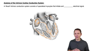
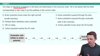
What are the key cellular features of cardiac muscle cells?
Cardiac muscle cells are short and branched, possess sarcomeres, have more mitochondria, less Ca2+, contain intercalated disks, and exhibit automaticity and autorhythmicity.
Name the three layers of the heart wall.
The three layers are the epicardium, myocardium, and endocardium.
What structures make up the pericardium?
The pericardium consists of the parietal and visceral layers, with the pericardial cavity containing pericardial fluid between them.
What are the main circuits of the circulatory system?
The main circuits are the pulmonary circuit, systemic circuit, and coronary circulation.
What are the primary types of blood vessels in the circulatory system?
The primary types are arteries, veins, and capillaries.
What are the four chambers of the heart?
The heart has two atria (right and left) and two ventricles (right and left).
What is the function of the interatrial septum?
The interatrial septum separates the right and left atria and contains the fossa ovalis, a remnant of the fetal foramen ovale.
What separates the ventricles of the heart?
The interventricular septum separates the right and left ventricles.
What are the atrioventricular (AV) valves and their function?
The AV valves (tricuspid on the right, bicuspid/mitral on the left) prevent backflow of blood from ventricles to atria during ventricular contraction.
Name the major veins entering the right atrium.
The superior vena cava, inferior vena cava, and coronary sinus enter the right atrium.
What valve controls blood flow from the right atrium to the right ventricle?
The right AV valve, also called the tricuspid valve, controls this flow.
Which valve leads blood from the right ventricle to the pulmonary trunk?
The pulmonary (semilunar) valve controls blood flow into the pulmonary trunk.
Which veins bring oxygenated blood to the left atrium?
The pulmonary veins bring oxygenated blood to the left atrium.
What valve controls blood flow from the left atrium to the left ventricle?
The left AV valve, also known as the bicuspid or mitral valve, controls this flow.
Which valve controls blood flow from the left ventricle to the aorta?
The aortic (semilunar) valve controls blood flow into the ascending aorta.
Name the major arteries branching from the aorta relevant to the heart.
The coronary arteries branch from the ascending aorta; the aorta also has an arch and a descending portion.
What is the ductus arteriosus and its adult remnant?
The ductus arteriosus is a fetal blood vessel connecting the pulmonary trunk to the aorta; it becomes the ligamentum arteriosum after birth.
What is the role of intercalated disks in cardiac muscle?
Intercalated disks connect cardiac muscle cells, allowing rapid electrical signal transmission and synchronized contraction.
What does automaticity in cardiac cells mean?
Automaticity is the ability of cardiac cells to generate their own electrical impulses without external stimuli.
What is the significance of more mitochondria in cardiac muscle cells?
More mitochondria provide the energy needed for continuous, rhythmic contractions of the heart muscle.
 Indietro
 6:13