 Indietro
Anatomy & Physiology: Heart and Blood Vessels Study Guide
 6:13

Termini in questo insieme (52)
The heart is located in the thoracic cavity, in the mediastinum, between the lungs, behind the sternum, and above the diaphragm, mostly left of the body's midline.
Apex: pointed inferior portion formed mainly by the left ventricle.
Base: broad superior/posterior portion where great vessels attach.
Pericardium: sac surrounding and protecting the heart.
Pericardial fluid: reduces friction between serous pericardial membranes during heartbeats.
Cardiac muscle is striated, involuntary, short, branched, usually with one nucleus, and connected by intercalated discs.
Intercalated discs: connect cardiac cells.
Desmosomes: mechanically hold cells together.
Gap junctions: allow rapid electrical signal passage.
Atria: thin-walled chambers receiving blood.
Auricles: muscular extensions increasing atrial capacity.
Ventricles: thick-walled chambers pumping blood out.
Interatrial septum: separates right and left atria.
Interventricular septum: separates ventricles and contains conduction pathway parts.
Trabeculae carneae: ridges in ventricles aiding contraction and preventing wall sticking.
Foramen ovale: fetal opening in interatrial septum allowing blood to bypass lungs.
Mitral valve: prevents backflow from left ventricle to left atrium.
Tricuspid valve: prevents backflow from right ventricle to right atrium.
Chordae tendineae attach AV valve cusps to papillary muscles.
Papillary muscles contract during systole to tighten chordae and prevent valve prolapse.
Valves open when pressure behind them is greater than ahead; they close when pressure ahead is greater, operating by pressure differences.
Rheumatic fever: inflammatory disease post-strep infection damaging heart valves.
Heart murmur: abnormal sound from turbulent blood flow, often due to valve issues.
To heart: superior/inferior vena cava, coronary sinus, pulmonary veins.
Away from heart: pulmonary trunk/arteries, aorta.
Right and left coronary arteries supply oxygenated blood; cardiac veins remove deoxygenated blood, draining into coronary sinus.
Pulmonary: RV → lungs → LA for gas exchange.
Systemic: LV → body → RA delivering oxygen and returning deoxygenated blood.
Coronary sulcus (atrioventricular groove), anterior interventricular sulcus, posterior interventricular sulcus; all mark chamber boundaries and contain vessels.
Specialized cardiac cells that spontaneously generate action potentials to establish and conduct the heart's electrical rhythm.
Cardiac muscle cells that contract to generate force needed to pump blood.
Ca²⁺: triggers contraction during plateau phase.
Na⁺: involved in depolarization.
K⁺: moves out causing repolarization.
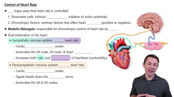
Epinephrine: increases heart rate and contractility.
Calcium-channel blockers: reduce Ca²⁺ entry, lowering heart rate and/or contractility.
Beta blockers: decrease heart rate and contractility by blocking sympathetic stimulation.
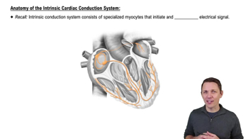
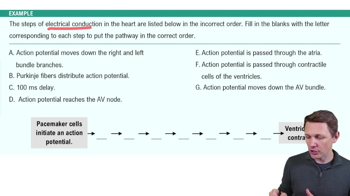
SA node → AV node → AV bundle (His) → right & left bundle branches → Purkinje fibers; SA node initiates impulse, AV node delays it.
Cardiac output is the blood volume pumped by one ventricle per minute.
Formula: CO = HR × SV
Systole: contraction phase.
Diastole: relaxation phase.
1. Ventricular filling
2. Atrial systole
3. Isovolumetric contraction
4. Ventricular ejection
5. Isovolumetric relaxation
P wave: atrial depolarization.
QRS complex: ventricular depolarization.
T wave: ventricular repolarization.
Normal sounds caused by valve closures (LUB = AV valves, DUB = semilunar valves).
Heart murmur caused by turbulent blood flow, often from abnormal valves.
SA node sets rate; autonomic nervous system modifies it: sympathetic increases HR, parasympathetic decreases HR.
EDV: blood volume in ventricle before contraction.
ESV: blood volume after contraction.
Stroke volume (SV): SV = EDV − ESV; affected by preload, contractility, afterload.
Cardiac output = heart rate × stroke volume; increasing either increases cardiac output.
Condition where heart cannot pump effectively, causing blood backup and fluid congestion (edema).
Narrowing/blockage of coronary arteries due to plaque, reducing blood and oxygen to myocardium, risking ischemia or infarction.
Arteries → arterioles → capillaries → venules → veins
1. Tunica externa: outer connective tissue.
2. Tunica media: smooth muscle and elastic tissue.
3. Tunica interna (intima): endothelium lining.
Arteries have thicker walls, thicker tunica media, smaller lumen, no valves.
Veins have thinner walls, larger lumen, and valves.
The hollow interior space through which blood flows.
Arteries: elastic (largest), muscular (medium), arterioles (smallest).
Veins: venules (small), medium veins (with valves), large veins (near heart).
Capillaries have walls one endothelial cell thick; function as exchange vessels for gases, nutrients, and wastes.
Networks of capillaries providing large surface area for exchange between blood and tissues.
Rings of smooth muscle controlling blood flow into capillary beds by contracting or relaxing.
Connections between blood vessels providing alternate routes for blood flow.
Valves prevent backflow of blood in veins.
Varicose veins are enlarged veins caused by valve failure and blood pooling.
Cardiac output, blood volume, peripheral resistance, blood viscosity, vessel diameter, and vessel elasticity.
Flow = Pressure difference ÷ Resistance; ↑ pressure or ↓ resistance increases flow.
Diffusion: movement of substances from high to low concentration.
Filtration: fluid pushed out of capillaries.
Reabsorption: fluid moves back into capillaries.
Blood hydrostatic pressure pushes fluid out; blood colloid osmotic pressure pulls fluid in.
Located in the medulla oblongata; regulates heart rate, contraction force, and vessel diameter to control blood pressure.
Nervous: autonomic changes in HR, contractility, vessel diameter.
Hormonal: hormones like epinephrine, ADH affect volume and resistance.
Autoregulation: local tissue control of blood flow.
Shock: inadequate tissue perfusion.
Hypovolemic shock: shock from blood/fluid loss.
Edema: excess fluid in tissues caused by pressure or protein imbalances.
Pulse: pressure wave from ventricular contraction.
Tachycardia: HR > 100 bpm.
Bradycardia: HR < 60 bpm.
Systolic BP: max pressure during ventricular contraction.
Diastolic BP: min pressure during relaxation.
Sphygmomanometer: device to measure BP.
Persistent high blood pressure.
Types: primary (no single cause), secondary (due to condition).
Lifestyle: exercise, healthy weight, low sodium, no smoking.
Diuretics: reduce blood volume.
Beta blockers: reduce heart rate and contractility.
Vasodilators/CCBs: relax vessels to lower resistance.