 Indietro
Anatomy & Physiology: Integumentary System and Skin Structure
 06:54

Termini in questo insieme (35)
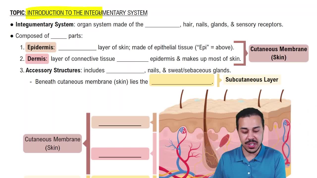
The superficial layer of skin made of stratified squamous keratinized epithelium. It is avascular and relies on diffusion from the dermis for oxygen and nutrients.
The deeper skin layer composed of loose and dense irregular collagenous connective tissue, including the papillary and reticular layers.
Also called subcutaneous tissue, it is the layer of adipose tissue beneath the skin.
Glands producing watery sweat for thermoregulation. They are the most numerous and active throughout life.
Located in axillae, anal area, and areola; secrete thick, protein-rich sweat active after puberty. Bacterial breakdown causes odor.
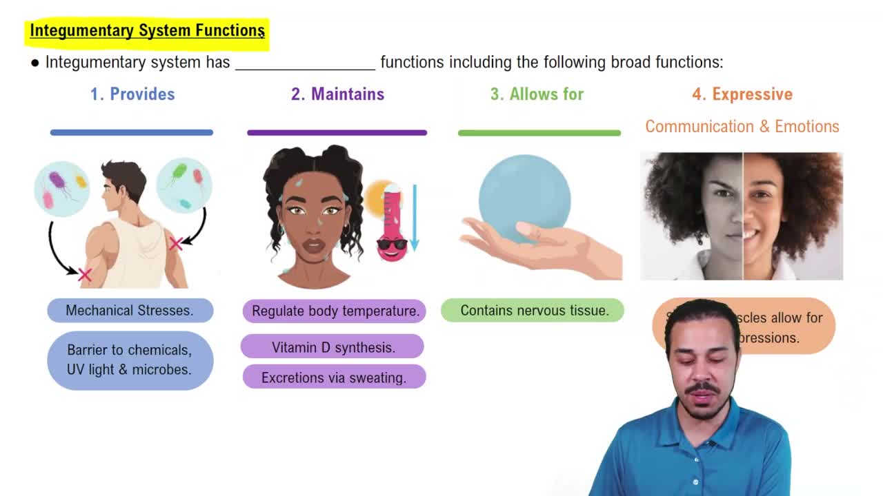
Protection, sensation, thermoregulation, excretion, and synthesis of vitamin D for calcium absorption.
If too hot, thermoreceptors signal hypothalamus to induce sweating and vasodilation; if too cold, vasoconstriction and blood redirection conserve heat.
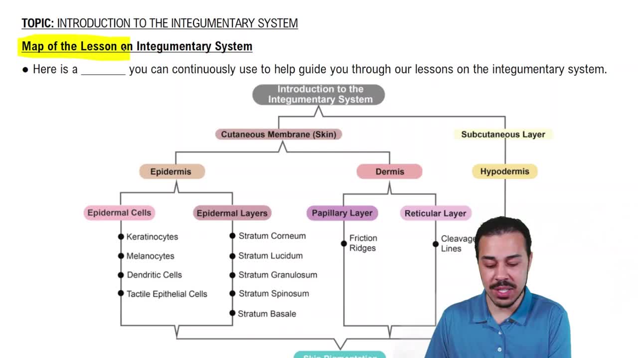
Stratum corneum, stratum lucidum (only in thick skin), stratum granulosum, stratum spinosum, stratum basale. Mnemonic: Come Let’s Get Sun Burned.
Make up 95% of epidermis; produce keratin which protects against mechanical damage and water loss.
Lowest epidermal layer where most mitosis occurs.
The thickest epidermal layer.
Layer where cells fill with keratin.
Clear layer found only in thick skin (palms and soles).
Top epidermal layer; cells take 40-50 days to migrate here.
Immune system phagocytes that protect skin and deeper tissues.
Oval cells in stratum basale that detect light touch and texture.
Cells that produce melanin, the pigment protecting against UV radiation.
Thickened area of stratum corneum formed by repetitive pressure.
Papillary layer is superficial with loose connective tissue; reticular layer is deeper with dense irregular connective tissue.
Sensory receptors in reticular dermis detecting pressure and vibration; resemble onion layers.
Folded epidermal patterns in thick skin that improve grip and form fingerprints.
Brown-black to orange-red pigment from melanocytes that protects skin from UV radiation.
Orange pigment found in foods like carrots and sweet potatoes.
Iron-containing red pigment in red blood cells that binds oxygen.
Blue skin color due to lack of oxygen.
Redness from increased blood flow in the dermis, such as during exercise or fever.
Loss of skin color due to decreased blood flow in the dermis.
Hair shaft projects above skin; root is embedded in follicle within dermis.
Smooth muscle that contracts to make hair stand on end (goosebumps).
Lanugo: fetal, thin, non-pigmented; Vellus: thin, non-pigmented; Terminal: thick, coarse, pigmented hair replacing vellus at puberty.
Nail plate rests on nail bed; nail body is visible part; eponychium is cuticle; hyponychium anchors nail plate distally; lunula is half-moon proximal region.
First degree: red, painful, no permanent damage; Second degree: partial thickness, blisters, may scar; Third degree: full thickness, often requires grafts.
Body divided into 11 sections of 9% each plus 1% perineum to estimate burn severity and guide treatment.
Basal cell carcinoma: least metastatic; Squamous cell carcinoma: keratinocyte cancer, more metastatic; Malignant melanoma: melanocyte cancer, most metastatic.
A: Asymmetry, B: Border irregularity, C: Color variation, D: Diameter, E: Evolving changes in mole.