Skip to main content
Anatomy & Physiology
Il mio corso
Impara
Preparazione agli esami
AI Tutor
Guide di studio
Soluzioni per libri di testo
Flashcard
Esplora
Prova l'app
Il mio corso
Impara
Preparazione agli esami
AI Tutor
Guide di studio
Soluzioni per libri di testo
Flashcard
Esplora
Prova l'app
Indietro
Anatomy & Physiology: Muscle and Joint Basics
Puoi toccare per girare la carta.
Types of joints and their mobility
Puoi toccare per girare la carta.
👆
Types of joints and their mobility
Fibrous
(immovable),
Cartilaginous
(slightly movable),
Synovial
(freely movable)
Avanzamenti del tracciato
I pulsanti di controllo sono stati cambiati in modalità "navigazione".
1/34
Video consigliati
VideoThumbView.guidedCourse
04:06
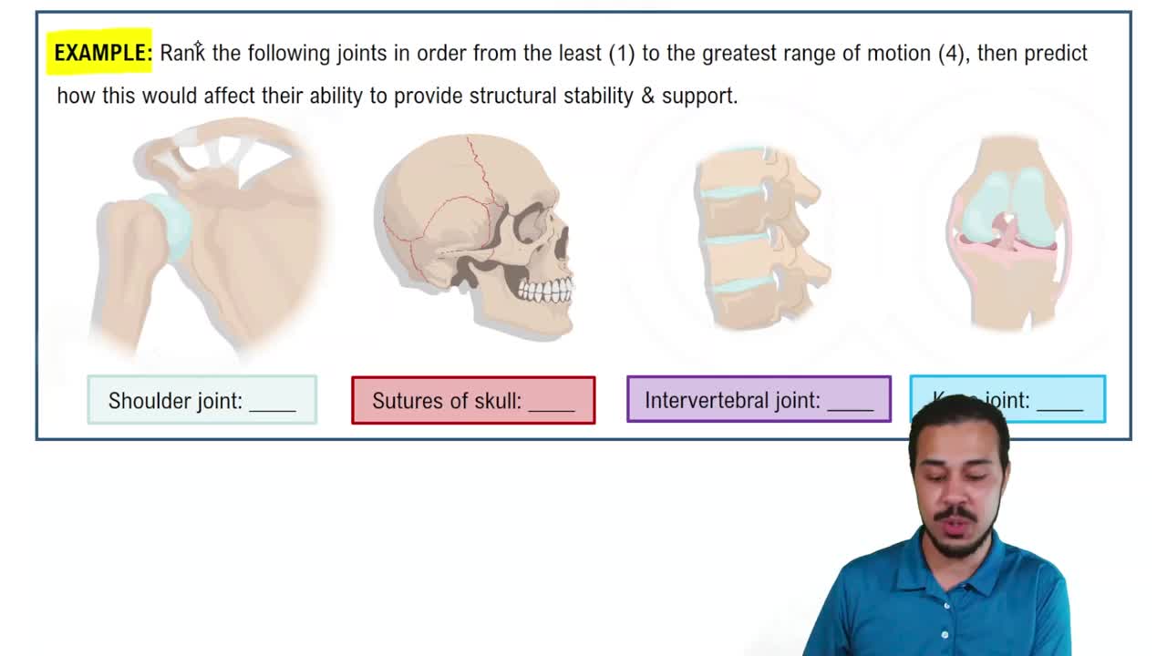
Introduction to Joints Example 1
11843
views
268
rank
VideoThumbView.guidedCourse
05:29
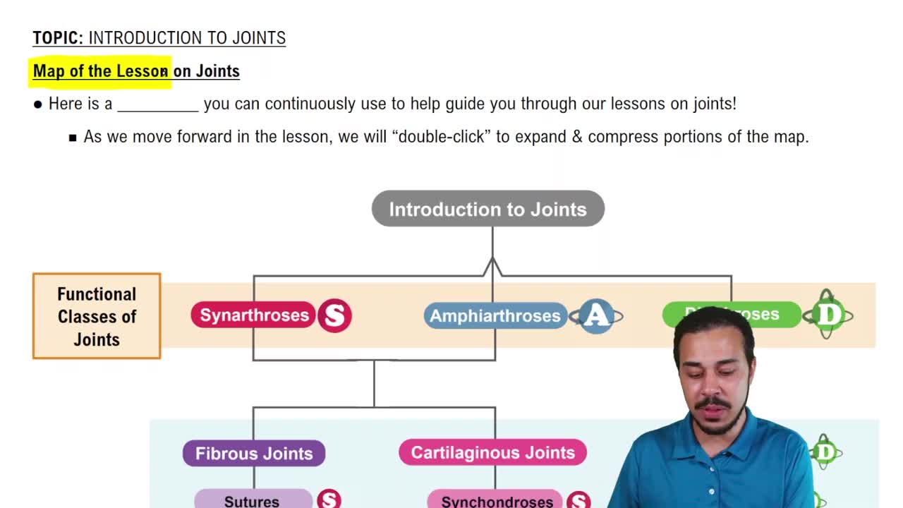
Map of the Lesson on Joints
12366
views
261
rank
1
comments
VideoThumbView.guidedCourse
05:39
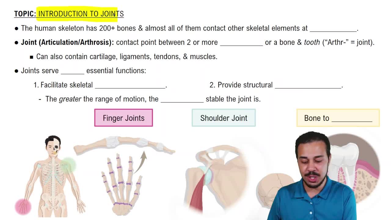
Introduction to Joints
16601
views
426
rank
Termini in questo insieme (34)
Nascondere definizioni
Types of joints and their mobility
Fibrous
(immovable),
Cartilaginous
(slightly movable),
Synovial
(freely movable)
Protein NOT in thin filament
Myosin
does not belong in a thin filament; it is part of the thick filament.
Sliding filament mechanism description
Thin filaments slide past thick filaments towards the
M line
during muscle contraction.
Key function in excitation-contraction coupling
The
storage and release of calcium ions
triggers muscle contraction.
Basic function of all muscle tissue
To
contract (shorten)
, generate tension, force, and produce movement.
Role of elastic fibers in muscle tissue
They resist excessive stretching and help muscle fibers
spring back
to original length after stretch.
Definition of a motor unit
A single motor neuron and all the skeletal muscle fibers it innervates.
Common features of skeletal and smooth muscle contractions
Both use
thick and thin filaments
, require
calcium
, and use
ATP
for energy.
What anchors thin and elastic filaments in myofibrils?
The
Z disc
(or Z line) anchors thin and elastic filaments.
Muscle response to physical inactivity
Muscle atrophy
, a decrease in muscle size and strength.
Muscles used in ventilation (breathing)
The
diaphragm
,
external intercostals
, and muscles for quiet exhalation.
Prime mover of elbow flexion
The
brachialis
muscle is the prime mover for elbow flexion.
Muscle group that flexes the leg at the knee when climbing stairs
The
hamstring group
flexes the leg at the knee.
Functions of the gluteus muscles
Gluteus Maximus
: extends thigh backward;
Gluteus Medius
: hip abduction;
Gluteus Minimus
: hip abduction and thigh inward rotation.
Muscles responsible for rotating thigh and pelvis during walking
Gluteus Medius
and
Gluteus Minimus
rotate the thigh and pelvis.
Agonist muscle function
The
prime mover
that contracts and shortens to produce a specific movement.
Antagonist muscle function
Opposes the agonist by gently lengthening and relaxing to control or stop motion.
Synergist muscle function
Assists the agonist by adding extra force during movement.
Fixator muscle function
Stabilizes the origin of the agonist so all pulling power moves the target bone.
Muscles of the abdomen and their fiber directions
Rectus abdominis
(vertical),
pyramidalis
(vertical),
external oblique
(inferomedial),
internal oblique
(superomedial),
transversus abdominis
(transverse).
Muscle that flexes and abducts the hand
The
flexor carpi radialis
flexes and abducts the hand.
Six levels of skeletal muscle structure
Whole muscle (organ), muscle fibers (cells), myofibrils, sarcomeres, myofilaments.
Location and description of the platysma muscle
A broad, thin sheet of muscle in the front triangle of the neck.
Angular movement moving a body part away from midline
Abduction
moves a body part away from the midline.
Radial collateral ligament (RCL) of the elbow
A short, strong band on the lateral (outer) side of the elbow.
Common locations of hinge joints
Found in knees, elbows, fingers, toes, and ankles.
Movement of forearm and hand toward face at elbow joint
Flexion
at the elbow joint.
Posterior and anterior cruciate ligaments of the knee
PCL
is at the back of the knee;
ACL
is at the front of the knee.
Structure of a synovial joint
A fluid-filled space called the
joint cavity
between articulating bones.
Primary motions of elbow and knee joints
Flexion
and
extension
are the main motions.
Function of tendon sheaths
Protective, fluid-filled layers that wrap around tendons to help smooth movement.
Joint movement when shaking head 'no'
Rotation
is the movement involved.
Example of abduction movement
Lifting the arm straight out to the side at the shoulder.
Annular ligament function
Strong band wrapping around the head of the radius, holding it against the ulna at the elbow.
 IndietroIndietro
 04:06