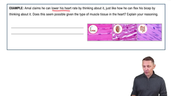
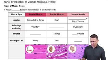
Skeletal: voluntary, striated, attached to bones; Cardiac: involuntary, striated, heart muscle with intercalated discs; Smooth: involuntary, non-striated, found in walls of organs.
Intercalated discs
Specialized connections between cardiac muscle cells that allow synchronized contraction by facilitating electrical and mechanical coupling.
Five common properties of muscle tissue
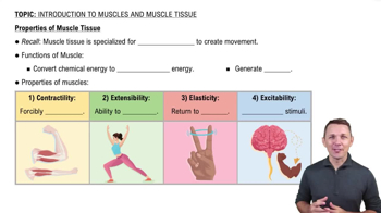
Contractility, Excitability, Conductivity, Distensibility, and Elasticity.
Sarcoplasm
The cytoplasm of a muscle cell containing glycogen and myoglobin for energy and oxygen storage.
Sarcolemma
The plasma membrane of a muscle fiber that conducts electrical impulses.
Myofibrils
Long, cylindrical organelles composed of myofilaments responsible for muscle contraction.
Sarcoplasmic reticulum
Specialized smooth ER in muscle cells that stores and releases calcium ions.
Transverse (T) tubules
Invaginations of the sarcolemma that transmit action potentials into the muscle fiber.
Triad in muscle cells
A structure formed by a T tubule flanked by two terminal cisternae of the sarcoplasmic reticulum.
Contractile proteins in muscle
Myosin (thick filament) and actin (thin filament) generate force during contraction.
Regulatory proteins in muscle
Tropomyosin and troponin control the interaction between actin and myosin.
Structural proteins in muscle
Proteins like titin and nebulin maintain the alignment and elasticity of myofilaments.
Sarcomere regions and structures
I band: thin filaments only; A band: thick and thin filaments overlap; H zone: thick filaments only; M line: center of thick filaments; Z disc: boundary of sarcomere.
Sliding filament mechanism
During contraction, thin filaments slide past thick filaments, shortening the sarcomere without changing filament length.
Changes in sarcomere during contraction
H zone shortens, Z discs move closer, A band length remains constant.
Resting membrane potential
The electrical gradient across the membrane of a resting cell, typically around -70 mV, due to ion distribution.
Difference between leak and gated ion channels
Leak channels are always open; gated channels open or close in response to stimuli.
Difference between ligand-gated and voltage-gated ion channels
Ligand-gated channels open when a chemical binds; voltage-gated channels open in response to changes in membrane potential.
Sodium-potassium pump function
Moves 3 Na+ ions out and 2 K+ ions into the cell using ATP to maintain resting potential.
 Indietro
 6:31