 Indietro
Anatomy & Physiology: Nervous System and Special Senses
 03:35

Termini in questo insieme (30)
Action potential is a rapid, temporary change in a neuron's membrane potential that propagates a nerve impulse along the axon.
Astrocytes are CNS glial cells that support neurons, maintain the blood-brain barrier, and regulate the extracellular environment.
The myelin sheath is a fatty layer that insulates axons, increasing the speed of nerve impulse conduction.
Nodes of Ranvier are gaps in the myelin sheath where ion exchange occurs, enabling saltatory conduction.
Saltatory conduction is the rapid transmission of action potentials jumping between nodes of Ranvier along myelinated axons.
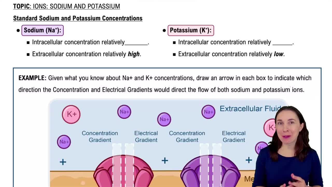
The resting membrane potential is the electrical charge difference across the membrane of a resting neuron, typically around -70 mV.
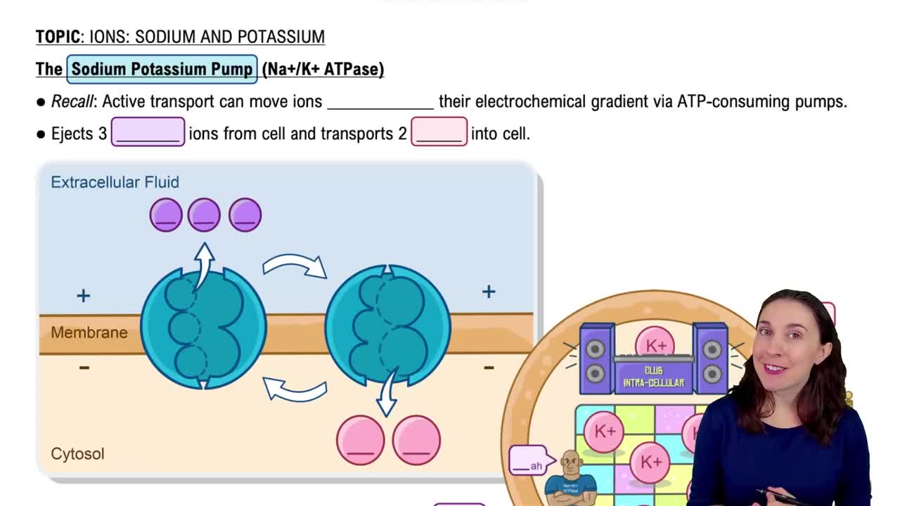
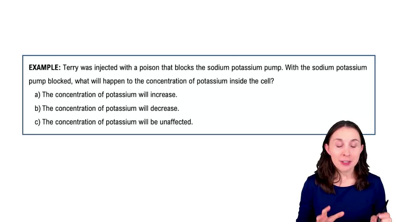
The sodium-potassium pump actively transports 3 Na+ out and 2 K+ into the neuron, maintaining resting potential.
Depolarization is the reduction of membrane potential, making the inside of the neuron less negative and triggering an action potential.
Repolarization restores the membrane potential to a negative value after depolarization during an action potential.
Hyperpolarization is when the membrane potential becomes more negative than the resting potential, inhibiting action potential generation.
Neurotransmitters are chemical messengers released at synapses to transmit signals between neurons or to effectors.
The CNS consists of the brain and spinal cord, responsible for processing and integrating information.
The PNS includes all nerves outside the CNS, connecting the CNS to limbs and organs.
The autonomic nervous system controls involuntary functions and regulates smooth muscle, cardiac muscle, and glands.
The somatic nervous system controls voluntary movements by innervating skeletal muscles.
A synapse is the junction where a neuron communicates with another neuron or effector cell via neurotransmitters.
Oligodendrocytes are CNS glial cells that form myelin sheaths around multiple axons.
Schwann cells form myelin sheaths around axons in the PNS, aiding in nerve regeneration.
CSF cushions the brain and spinal cord, removes waste, and provides a stable chemical environment.
The blood-brain barrier protects the CNS by restricting passage of substances from blood to brain tissue.
The cerebrum is the largest brain part, responsible for higher brain functions like thought, memory, and voluntary movement.
Basal nuclei are clusters of neurons in the cerebrum involved in movement regulation.
The cerebellum coordinates voluntary movements, balance, and posture.
The thalamus acts as a relay station for sensory information to the cerebral cortex.
The hypothalamus regulates homeostasis, including temperature, hunger, and hormone release.
Photoreceptors are retinal cells (rods and cones) that detect light and color.
Rod cells detect low light levels and are responsible for night vision.
Cone cells detect color and function best in bright light.
Ossicles (malleus, incus, stapes) transmit sound vibrations from the eardrum to the inner ear.
Semicircular canals detect rotational movements for dynamic equilibrium.