Skip to main content
Anatomy & Physiology
Il mio corso
Impara
Preparazione agli esami
AI Tutor
Guide di studio
Soluzioni per libri di testo
Flashcard
Esplora
Prova l'app
Il mio corso
Impara
Preparazione agli esami
AI Tutor
Guide di studio
Soluzioni per libri di testo
Flashcard
Esplora
Prova l'app
Indietro
Anatomy & Physiology Practical Exam II Key Concepts
Puoi toccare per girare la carta.
Lymph node key features
Puoi toccare per girare la carta.
👆
Lymph node key features
Identify structures such as the cortex, medulla, lymphatic nodules, and germinal centers on lymph node slides or models.
Avanzamenti del tracciato
I pulsanti di controllo sono stati cambiati in modalità "navigazione".
1/20
Video consigliati
VideoThumbView.guidedCourse
3:32
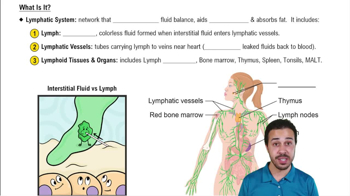
Introduction to the Lymphatic System Example 2
4847
views
88
rank
VideoThumbView.guidedCourse
3:33
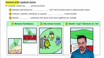
What Is It?
6484
views
167
rank
VideoThumbView.guidedCourse
3:57
Functions of the Lymphatic System
5496
views
128
rank
Termini in questo insieme (20)
Nascondere definizioni
Lymph node key features
Identify structures such as the cortex, medulla, lymphatic nodules, and germinal centers on lymph node slides or models.
Spleen key features
Recognize white pulp, red pulp, and trabeculae on spleen slides or models.
Primary organs of the lymphatic system
Include lymph nodes, spleen, thymus, and mucosa-associated lymphoid tissue (MALT).
Function of the thymus
Site of T cell maturation and development in the immune system.
Role of MALT
Protects mucosal surfaces by producing immune responses to pathogens at mucous membranes.
Antigens vs. antibodies
Antigens are foreign molecules that trigger immune responses; antibodies are proteins that specifically bind antigens.
Classes of antibodies
IgG, IgA, IgM, IgE, and IgD differ in structure and immune function.
Three tunics of blood vessels
Tunica intima, tunica media, and tunica externa (adventitia) compose arteries and veins.
Differences between arteries and veins
Arteries have thicker tunica media and smaller lumen; veins have valves and larger lumen.
Naming of arteries
Usually named for the region they supply or the organ they serve, e.g., subclavian or inferior mesenteric.
Hepatic portal system function
Transports nutrient-rich blood from the digestive organs to the liver for processing.
Fetal circulation shunts
Include the ductus arteriosus, foramen ovale, and ductus venosus, which bypass nonfunctional fetal organs.
Postnatal fate of fetal shunts
Ductus arteriosus becomes ligamentum arteriosum; foramen ovale closes forming fossa ovalis; ductus venosus becomes ligamentum venosum.
Layers of the trachea
Mucosa, submucosa, cartilaginous layer (with trachealis muscle), and adventitia.
Histological features of tracheal mucosa
Contains ciliated pseudostratified columnar epithelium and goblet cells.
Histology of lung tissue
Includes alveoli lined by simple squamous epithelium and rich capillary networks.
Muscles of quiet breathing
Diaphragm and external intercostal muscles.
Muscles of forced breathing
Include sternocleidomastoid, scalenes, pectoralis minor, and internal intercostals.
Respiratory volumes and capacities
Include tidal volume, inspiratory reserve volume, expiratory reserve volume, vital capacity, and total lung capacity.
Spirometer readout interpretation
Used to measure lung volumes and capacities to assess respiratory function.
 IndietroIndietro
 3:32
3:32