 Indietro
Anatomy & Physiology: The Heart - Chapter 19 Study Guide
 6:13

Termini in questo insieme (26)
The right side receives oxygen-poor blood and pumps it to the lungs (pulmonary circuit). The left side receives oxygen-rich blood from the lungs and pumps it to the body (systemic circuit).
The heart lies in the mediastinum between the lungs. The apex points inferiorly and to the left, and the base is the broad posterior surface.
Fibrous pericardium → parietal serous pericardium → pericardial cavity → epicardium → myocardium → endocardium.
Strong outer layer of dense connective tissue that protects and anchors the heart.
Cardiac muscle layer arranged in circular and spiral patterns; generates the pumping force of the heart.
Right and left atria: superior receiving chambers. Right and left ventricles: inferior pumping chambers.
Interatrial septum separates the atria; interventricular septum separates the ventricles.
External groove separating atria from ventricles; contains coronary blood vessels.
Receives oxygen-poor blood from superior vena cava, inferior vena cava, and coronary sinus; contains pectinate muscles, crista terminalis, fossa ovalis, and tricuspid valve.
Receives blood from right atrium via tricuspid valve; pumps blood through pulmonary valve to pulmonary trunk; contains trabeculae carneae, papillary muscles, and chordae tendineae.
Receives oxygen-rich blood from four pulmonary veins; blood passes through mitral (bicuspid) valve to left ventricle.
Receives blood through mitral valve; pumps through aortic valve into aorta; has thick walls and contains trabeculae carneae, papillary muscles, and chordae tendineae; forms the apex of the heart.
Chordae tendineae connect papillary muscles to AV valve cusps, preventing valve eversion during ventricular contraction; papillary muscles contract to tighten chordae tendineae.
AV valves are between atria and ventricles and open when atrial pressure exceeds ventricular pressure. Semilunar valves are between ventricles and arteries and open when ventricular pressure exceeds arterial pressure.
S1 ('lub') is caused by closure of AV valves; S2 ('dup') is caused by closure of semilunar valves.
Blood flows: venae cavae → right atrium → tricuspid valve → right ventricle → pulmonary valve → pulmonary trunk → lungs → pulmonary veins → left atrium → mitral valve → left ventricle → aortic valve → aorta → body.
The left ventricle pumps blood into the systemic circuit, which has higher resistance and longer distance, requiring a thicker muscular wall.
Specialized junctions connecting cardiac muscle cells; contain fasciae adherens and desmosomes for mechanical binding, and gap junctions for electrical coupling.
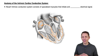
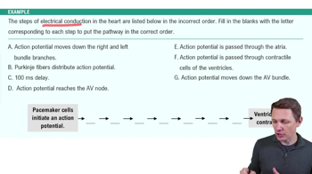
SA node → internodal pathways → AV node → AV bundle → right and left bundle branches → Purkinje fibers.
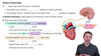
Parasympathetic (vagus nerve) decreases heart rate; sympathetic increases heart rate and contraction strength.
Right coronary artery supplies right and posterior heart; left coronary artery branches into anterior interventricular (LAD) and circumflex arteries supplying left and anterior heart.
Atherosclerosis reduces coronary blood flow; angina causes chest pain; myocardial infarction causes cardiac muscle death; heart failure weakens pumping ability.
Ventricular septal defect causes blood mixing; transposition of great vessels separates circuits; coarctation of aorta narrows vessel increasing workload; tetralogy of Fallot combines four defects reducing pulmonary flow.
Valve thickening, reduced cardiac reserve, fibrosis replacing cardiac cells, and decreased maximum heart rate; exercise can improve heart health.
Pulmonary arteries carry oxygen-poor blood to lungs; pulmonary veins return oxygen-rich blood to the heart.
Atrial fibrillation causes irregular atrial impulses increasing stroke risk; ventricular fibrillation causes uncoordinated ventricular contractions leading to cardiac arrest.