 Indietro
Anatomy & Physiology: Tissue Structure and Function
 03:32

Termini in questo insieme (36)
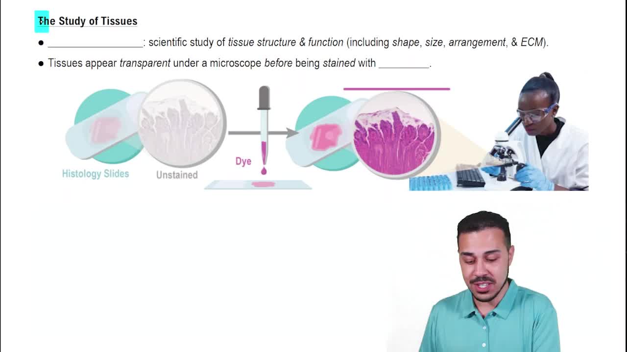
Histology is the study of tissues.
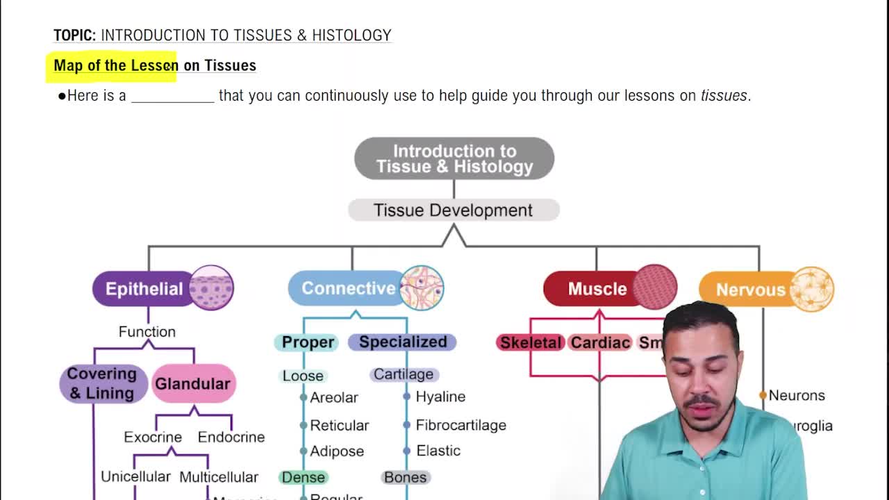
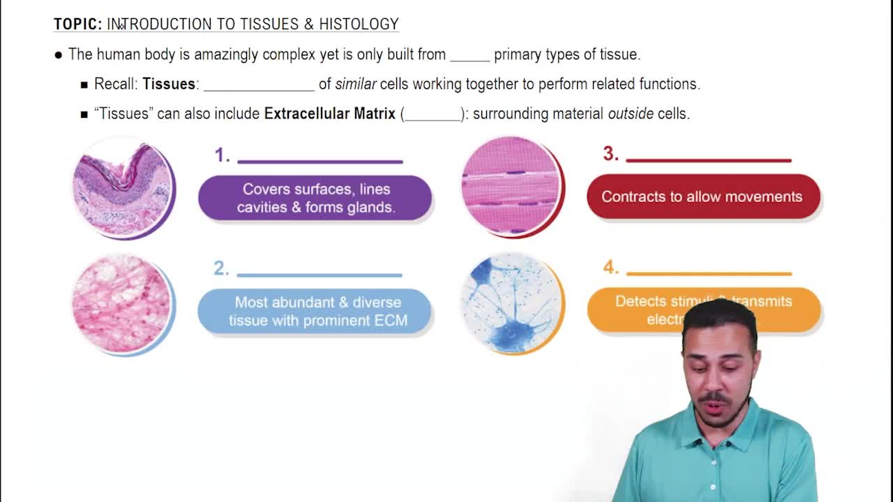
Epithelial, Connective, Muscle, and Nervous tissues.
Covers exposed surfaces, lines internal passageways, and forms glands.
Fills internal spaces, supports other tissues, transports materials, and stores energy.
Microvilli increase surface area where all absorption takes place.
Squamous (thin, flat), Cuboidal (square), and Columnar (tall, slender rectangles).
Simple epithelium (single layer) and Stratified epithelium (multiple layers).
Thin for absorption and diffusion; lines body cavities (mesothelium) and blood vessels (endothelium).
Protects against mechanical stresses; keratin adds strength and water resistance.
Simple squamous is in kidneys; stratified squamous is in glands.
Absorption and secretion; found in stomach, small and large intestines.
Appears layered but is single-layered; typically ciliated; found in nasal cavity, trachea, and bronchi.
Provides protection; rare; found in pharynx, anus, and urethra.
Fibroblasts (produce protein), Adipocytes (fat cells), Macrophages (phagocytic immune cells), Mast cells (inflammation), Lymphocytes, Microphages, and Mesenchymal stem cells.
Engulf pathogens and damaged cells; fixed macrophages stay in tissue, free macrophages migrate.
Stimulate inflammation by releasing histamine and heparin after injury or infection.
Collagen fibers: strong, resist force in one direction; Reticular fibers: form flexible networks; Elastic fibers: branched, return to original length after stretching.
Contains adipocytes that store fat; cells expand or shrink; mesenchymal cells produce new fat cells when needed.
White fat stores energy and insulates; brown fat is vascularized, rich in mitochondria, and generates heat in infants.
Tightly packed, parallel collagen fibers; found in tendons, ligaments, and aponeuroses.
Connective tissue layers that support and surround organs; include superficial, deep, and subserous fascia.
Plasma (watery matrix), red blood cells (erythrocytes), white blood cells (leukocytes), and platelets.
Interstitial fluid that enters lymphatic vessels, monitored by the immune system, and returned to veins near the heart.
Shock absorption and reducing friction between bones.
Hyaline: most common, tough, flexible; Elastic: supportive, bends easily; Fibrocartilage: very durable, limits movement.
Supports weight, is calcified for rigidity, resists shattering due to collagen fibers, and contains osteocytes in lacunae.
Physical barriers that line or cover body surfaces; consist of epithelium supported by connective tissue.
Mucous, Serous, Cutaneous, and Synovial membranes.
Skeletal muscle: large muscles for movement; Cardiac muscle: heart only; Smooth muscle: walls of hollow organs.
Conducting electrical impulses; concentrated in brain and spinal cord.
Neurons (signal transmission) and Neuroglia (supporting cells).
Trauma or infection; damaged cells release prostaglandins, proteins, and potassium ions; mast cells activate inflammation.
Tissue destruction after injury; necrotic tissue and pus accumulate, sometimes forming an abscess.
Epithelia, connective tissues (except cartilage), and smooth muscle regenerate well; skeletal muscle, cardiac muscle, and nervous tissue regenerate poorly or not at all.
Slower repair, thinner epithelia, fragile connective tissue, brittle bones, increased bruising, cardiovascular disease, and mental deterioration.
Cancer rates increase with age; 25% of people develop cancer; most caused by chemical or environmental factors; 40% linked to cigarette smoke.