 Indietro
Blood Vessels - Anatomy & Physiology
 3:38

Termini in questo insieme (28)
Tunica intima, tunica media, and tunica externa.
Provides a smooth, low-friction surface next to the blood, composed of endothelium.
Contains circular smooth muscle that controls vessel diameter via vasoconstriction and vasodilation; it is the thickest layer in arteries.
Protects, strengthens, anchors the vessel, and nourishes the outer wall; contains longitudinal connective tissue fibers and vasa vasorum in large vessels.
The central, blood-filled space inside a vessel.
Largest conducting arteries with abundant elastin; they smooth the pressure surge from ventricular ejection (e.g., aorta, pulmonary trunk).
Muscular arteries are distributing arteries with a thick tunica media and distinct elastic membranes; they supply most named arteries.
Smallest arteries that regulate blood flow into capillary beds by changing diameter in response to local and sympathetic signals.
Vasoconstriction: smooth muscle contracts, narrowing lumen.
Vasodilation: smooth muscle relaxes, widening lumen.
Continuous: least permeable, tight junctions.
Fenestrated: pores for rapid exchange.
Sinusoid: wide gaps, very leaky for large molecules and cells.
Most organs; special tight form creates the blood–brain barrier.
They stabilize capillaries, regulate permeability, and contribute to new vessel formation.
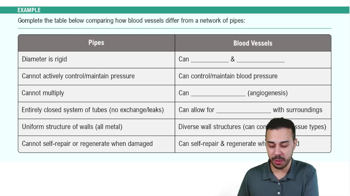
Veins have thinner walls, less elastin, larger lumens, and a relatively thick tunica externa; they carry blood toward the heart at lower pressure.
Venous valves prevent backflow, skeletal muscle pump compresses veins, and respiratory pump uses breathing pressure changes.
Pulmonary: right ventricle to lungs, lower pressure.
Systemic: left ventricle to body tissues, higher pressure.
A connection between neighboring arteries providing collateral routes around blockages, common near joints and in the heart and brain.
Brachiocephalic trunk, left common carotid artery, left subclavian artery.
Subclavian → axillary → brachial → radial and ulnar → palmar arches → digital arteries.
A portal system connecting two capillary beds: GI tract capillaries absorb nutrients, then blood passes through liver sinusoids for processing before returning to the heart.
Foramen ovale → fossa ovalis; ductus arteriosus → ligamentum arteriosum; ductus venosus → ligamentum venosum.
Because vessel names reflect direction relative to the heart: pulmonary arteries leave the heart; pulmonary veins return to it.
Shunts blood from the right atrium to the left atrium, bypassing the non-functioning fetal lungs.
First breaths expand lungs, lowering pulmonary resistance; increased left atrial pressure closes foramen ovale; ductus arteriosus constricts; umbilical vessels close.
A localized ballooning or dilation of a weakened vessel wall that can rupture and cause life-threatening hemorrhage.
A clot in a deep vein, often in the lower limb; a detached clot can travel to the lungs causing pulmonary embolism.
Great saphenous empties into the femoral vein; small saphenous empties into the popliteal vein.
Postcapillary venules.
Small vessels that supply blood to the outer walls of large blood vessels.