 Indietro
Cardiovascular System: Heart - Anatomy & Physiology
 6:13

Termini in questo insieme (20)
The three layers are the epicardium (outer layer), myocardium (middle muscular layer), and endocardium (inner lining).
The pericardium is a double-layered sac that encloses the heart, providing protection and reducing friction during heartbeats.
The right atrium receives deoxygenated blood; the right ventricle pumps it to the lungs; the left atrium receives oxygenated blood from the lungs; the left ventricle pumps oxygenated blood to the body.
The tricuspid valve is between right atrium and ventricle; the pulmonary valve between right ventricle and pulmonary artery; the mitral (bicuspid) valve between left atrium and ventricle; the aortic valve between left ventricle and aorta.
Blood flows from the right ventricle through the pulmonary valve into the pulmonary arteries, to the lungs for oxygenation, then returns via pulmonary veins to the left atrium.
Oxygenated blood leaves the left ventricle through the aortic valve into the aorta, travels through body tissues, and returns deoxygenated blood to the right atrium via the vena cavae.
Coronary arteries supply oxygen-rich blood to the heart muscle itself, ensuring it receives nutrients and oxygen for contraction.
Cardiac muscle is involuntary, striated, has intercalated discs for synchronized contraction, and is resistant to fatigue, unlike skeletal muscle which is voluntary.
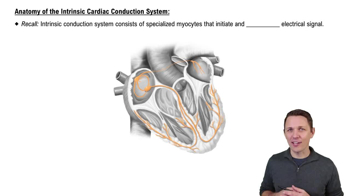
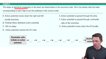
The system includes the sinoatrial (SA) node, atrioventricular (AV) node, bundle of His, bundle branches, and Purkinje fibers.
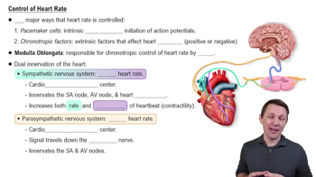
The SA node acts as the heart's natural pacemaker, initiating electrical impulses that trigger heartbeats.
An ECG records the electrical activity of the heart, helping diagnose arrhythmias, heart damage, and other cardiac conditions.
The cardiac cycle includes atrial systole, ventricular systole, and diastole (relaxation phase).
'Lub' is caused by the closing of the atrioventricular valves; 'dub' is caused by the closing of the semilunar valves.
Heart murmurs are abnormal sounds caused by turbulent blood flow, often due to valve defects or leaks.
Stroke volume is influenced by preload, contractility, and afterload.
The sympathetic nervous system increases heart rate and contractility; the parasympathetic system decreases heart rate.
Stroke volume is the amount of blood ejected by a ventricle with each heartbeat.
The AV node is located between the atria and ventricles; it delays the electrical impulse to allow ventricular filling before contraction.
Purkinje fibers rapidly conduct impulses throughout the ventricles, causing coordinated ventricular contraction.
The left ventricle has a thicker myocardium to pump blood at higher pressure through systemic circulation; the right ventricle pumps to the lungs at lower pressure.