 Indietro
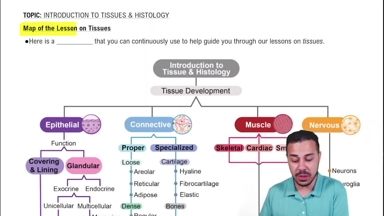
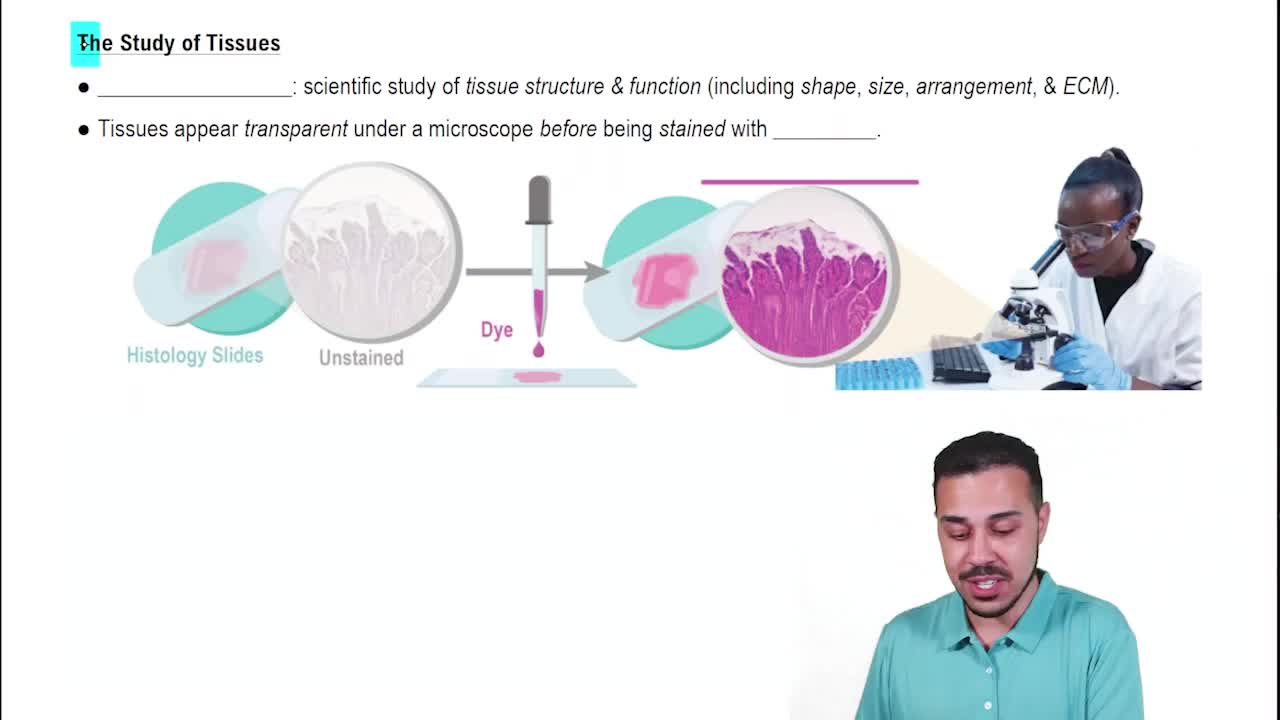
Epithelial and Connective Tissue Identification
 03:32
03:32
Termini in questo insieme (20)
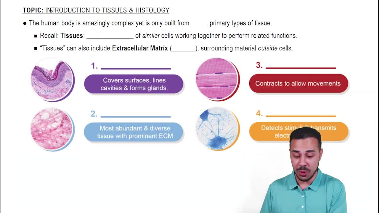
Epithelial tissue covers body surfaces, lines cavities, and forms glands. It has closely packed cells with minimal extracellular matrix and is avascular.
Squamous (flat), cuboidal (cube-shaped), and columnar (tall and column-like) cells.
Simple epithelium has a single cell layer; stratified epithelium has multiple layers.
Connective tissue supports, binds, and protects organs and tissues, and stores energy.
Cells, fibers, and ground substance (extracellular matrix).
Collagen fibers (strength), elastic fibers (stretch), and reticular fibers (support network).
Collagen fibers appear thick, wavy, and pinkish with H&E stain, providing tensile strength.
Elastic fibers are thin, dark-staining, and branched, allowing tissue to stretch and recoil.
Reticular fibers are thin, branching fibers forming a supportive mesh in soft organs like lymph nodes.
Dense regular connective tissue contains parallel collagen fibers and resists pulling forces.
Loose connective tissue has fewer fibers and more ground substance, appearing less dense and more flexible.
A thin layer of extracellular matrix that anchors epithelial tissue to underlying connective tissue.
Single layer of flat, thin cells with a centrally located nucleus, resembling floor tiles.
Cells appear layered due to nuclei at different levels but all cells contact the basement membrane; often ciliated.
Hyaline cartilage has a glassy matrix with chondrocytes in lacunae; dense connective tissue has densely packed collagen fibers without lacunae.
Fibroblasts are spindle-shaped cells that produce fibers and ground substance, often with elongated nuclei.
Adipose tissue stores fat; cells appear as large empty spaces (lipid droplets) with nuclei pushed to the side.
Stratified squamous epithelium has multiple layers with flat cells at the surface; simple squamous has only one layer.
Elastic fibers provide stretch and recoil in tissues like skin and lungs.
Provides structural support, mediates biochemical signals, and determines tissue properties.