 Indietro
Heart Anatomy - Anatomy & Physiology
 6:13

Termini in questo insieme (14)
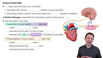
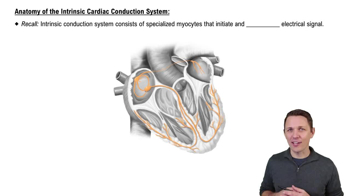
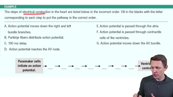
Pacemaker cells are specialized cardiac muscle cells that generate and conduct electrical impulses, initiating the heartbeat and regulating heart rhythm.
Desmosomes and gap junctions occur in cardiac muscle cells; desmosomes provide mechanical strength, and gap junctions allow electrical communication.
Desmosomes anchor cardiac muscle cells together, preventing them from pulling apart during contraction.
Gap junctions allow ions to pass directly between cardiac cells, enabling synchronized contraction.
The structure labeled 1 is the right atrium, which receives deoxygenated blood from the body.
The structure labeled 2 is the right ventricle, which pumps deoxygenated blood to the lungs.
The structure labeled 3 is the left atrium, which receives oxygenated blood from the lungs.
The structure labeled 4 is the left ventricle, which pumps oxygenated blood to the body.
The structure labeled 5 is the tricuspid valve, which prevents backflow of blood from the right ventricle to the right atrium.
The structure labeled 6 is the mitral (bicuspid) valve, which prevents backflow from the left ventricle to the left atrium.
The structure labeled 7 is the interventricular septum, which separates the right and left ventricles.
The structure labeled 8 is the aorta, which carries oxygenated blood from the left ventricle to the body.
The structure labeled 9 is the pulmonary artery, which carries deoxygenated blood from the right ventricle to the lungs.
The structure labeled 10 is the pulmonary veins, which return oxygenated blood from the lungs to the left atrium.