Skip to main content
Anatomy & Physiology
Il mio corso
Impara
Preparazione agli esami
AI Tutor
Guide di studio
Soluzioni per libri di testo
Flashcard
Esplora
Prova l'app
Il mio corso
Impara
Preparazione agli esami
AI Tutor
Guide di studio
Soluzioni per libri di testo
Flashcard
Esplora
Prova l'app
Indietro
Heart Anatomy and Circulatory System Histology
Puoi toccare per girare la carta.
Parietal pericardium
Puoi toccare per girare la carta.
👆
Parietal pericardium
The outer layer of the pericardium that lines the fibrous pericardium and surrounds the heart.
Avanzamenti del tracciato
I pulsanti di controllo sono stati cambiati in modalità "navigazione".
1/27
Video consigliati
6:13
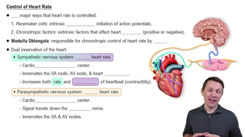
Control of Heart Rate
8097
views
167
rank
1
comments
8:41
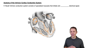
Anatomy of the Intrinsic Cardiac Conduction System
13835
views
338
rank
3
comments
3:42
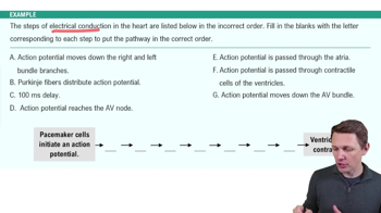
Electrical Conduction System of the Heart Example 2
7593
views
186
rank
Termini in questo insieme (27)
Nascondere definizioni
Parietal pericardium
The outer layer of the pericardium that lines the fibrous pericardium and surrounds the heart.
Visceral pericardium
Also called the epicardium, it is the inner layer of the pericardium that directly covers the heart muscle.
Pericardial cavity
The space between the parietal and visceral pericardium containing lubricating fluid to reduce friction during heartbeats.
Myocardium
The thick middle layer of the heart wall composed of cardiac muscle tissue responsible for contraction.
Endocardium
The innermost layer of the heart wall made of simple squamous endothelium lining the heart chambers.
Fibrous skeleton of the heart
A dense connective tissue framework that supports the heart valves and separates atria from ventricles.
Interventricular septum
The muscular wall separating the right and left ventricles of the heart.
Atrioventricular valves
Valves between atria and ventricles: the tricuspid valve on the right and the bicuspid (mitral) valve on the left.
Semilunar valves
Valves located at the bases of the pulmonary trunk and aorta: pulmonary semilunar valve and aortic semilunar valve.
Right coronary artery
Supplies blood to the right atrium, right ventricle, and parts of the conduction system; includes marginal and posterior interventricular arteries.
Left coronary artery
Supplies blood to the left atrium, left ventricle, and interventricular septum; includes anterior interventricular and circumflex arteries.
Great cardiac vein
Drains blood from the anterior heart and empties into the coronary sinus.
Superior vena cava
Large vein that returns deoxygenated blood from the upper body to the right atrium.
Ascending aorta
The initial section of the aorta that arises from the left ventricle and supplies systemic circulation.
Common carotid artery
Major artery supplying blood to the head and neck, dividing into internal and external carotid arteries.
Tunica media
The middle layer of blood vessels composed mainly of smooth muscle responsible for vasoconstriction and vasodilation.
Tunica interna (intima)
The innermost layer of blood vessels lined by simple squamous endothelium and includes the internal elastic lamina.
Purkinje fibers
Specialized cardiac muscle fibers that conduct electrical impulses rapidly through the ventricles.
Intercalated discs
Specialized connections between cardiac muscle cells that allow synchronized contraction.
Chordae tendineae
Tendinous cords that connect papillary muscles to atrioventricular valve leaflets, preventing valve prolapse.
Papillary muscles
Muscle projections from the ventricular walls that anchor chordae tendineae to prevent valve inversion.
Trabeculae carneae
Irregular muscular ridges on the inner walls of the ventricles that help with contraction efficiency.
Moderator band
A muscular band in the right ventricle (noted in sheep hearts) that carries part of the conduction system.
Azygos vein
A vein running along the thoracic vertebral column that drains blood from the thorax into the superior vena cava.
Hemiazygos vein
A vein that drains the lower left thoracic wall and empties into the azygos vein.
Internal jugular vein
Major vein draining blood from the brain, face, and neck into the brachiocephalic vein.
Simple squamous epithelium in circulatory system
The tissue type lining the endocardium and tunica interna, providing a smooth surface for blood flow.
 IndietroIndietro
 6:13