Skip to main content
Anatomy & Physiology
Il mio corso
Impara
Preparazione agli esami
AI Tutor
Guide di studio
Soluzioni per libri di testo
Flashcard
Esplora
Prova l'app
Il mio corso
Impara
Preparazione agli esami
AI Tutor
Guide di studio
Soluzioni per libri di testo
Flashcard
Esplora
Prova l'app
Indietro
Heart Anatomy and Physiology - Chapter 18
Puoi toccare per girare la carta.
Position of the heart in the thoracic cavity
Puoi toccare per girare la carta.
👆
Position of the heart in the thoracic cavity
The heart is located in the mediastinum, between the lungs, posterior to the sternum, and above the diaphragm.
Avanzamenti del tracciato
I pulsanti di controllo sono stati cambiati in modalità "navigazione".
1/26
Video consigliati
6:13
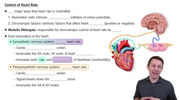
Control of Heart Rate
8023
views
166
rank
1
comments
8:41
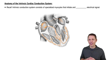
Anatomy of the Intrinsic Cardiac Conduction System
13670
views
335
rank
3
comments
3:42
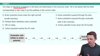
Electrical Conduction System of the Heart Example 2
7535
views
185
rank
Termini in questo insieme (26)
Nascondere definizioni
Position of the heart in the thoracic cavity
The heart is located in the mediastinum, between the lungs, posterior to the sternum, and above the diaphragm.
Four chambers of the heart
The heart has two atria (right and left) and two ventricles (right and left).
Coronary sulcus
A groove on the heart's surface that separates the atria from the ventricles and contains coronary blood vessels.
Anterior and posterior interventricular sulci
Grooves that mark the boundary between the right and left ventricles on the front and back of the heart.
Function of heart septa
The septa separate the right and left sides of the heart to prevent mixing of oxygen-rich and oxygen-poor blood.
Atrioventricular (AV) valves
Valves between atria and ventricles (tricuspid on right, mitral on left) that prevent backflow during ventricular contraction.
Semilunar valves
Valves (pulmonary and aortic) located at the exits of ventricles that prevent backflow of blood into the heart after ejection.
Layers of the pericardium
Includes the fibrous pericardium (outer tough layer) and serous pericardium (parietal and visceral layers) with the pericardial cavity between them.
Myocardium and intercalated discs
The myocardium is the thick cardiac muscle layer; intercalated discs connect cardiac cells and allow synchronized contraction.
Endocardium
A thin, smooth inner lining of the heart chambers that reduces friction for blood flow.
Right and left coronary arteries
Arteries that supply oxygen-rich blood to the heart muscle; they branch off the ascending aorta.
Cardiac veins and coronary sinus
Veins that collect oxygen-poor blood from the myocardium and drain into the coronary sinus, which empties into the right atrium.
Phases of cardiac muscle action potential
Depolarization (Na+ influx), plateau (Ca2+ influx balances K+ efflux), and repolarization (K+ efflux).
Action potential generation in pacemaker vs contractile cells
Pacemaker cells have spontaneous depolarization; contractile cells require stimulation and have a plateau phase; skeletal muscle cells lack plateau.
Significance of the plateau phase
Prolongs contraction to allow efficient blood ejection and prevents tetanus in cardiac muscle.
Major blood vessels entering and leaving the heart
Entering: superior/inferior vena cava (veins, oxygen-poor), pulmonary veins (veins, oxygen-rich). Leaving: pulmonary trunk (artery, oxygen-poor), aorta (artery, oxygen-rich).
Blood flow through the heart
Blood flows: vena cava → right atrium → tricuspid valve → right ventricle → pulmonary valve → pulmonary trunk → lungs → pulmonary veins → left atrium → mitral valve → left ventricle → aortic valve → aorta.
Heart as a double pump
The right side pumps blood to the lungs (pulmonary circuit), and the left side pumps blood to the body (systemic circuit).
Parts of the cardiac conduction system
Includes the SA node, AV node, AV bundle (Bundle of His), bundle branches, and Purkinje fibers.
SA node pacing role
The SA node is the primary pacemaker due to its fastest spontaneous depolarization rate, setting the heart rhythm.
Autonomic nervous system role in cardiac function
The ANS modulates heart rate and force of contraction via sympathetic (increases) and parasympathetic (decreases) inputs.
Waveforms in a normal ECG
P wave (atrial depolarization), QRS complex (ventricular depolarization), T wave (ventricular repolarization).
ECG waveforms and cardiac events
P wave: atrial contraction; QRS: ventricular contraction; T wave: ventricular relaxation.
Definition of cardiac cycle, systole, and diastole
Cardiac cycle: one complete heartbeat; systole: contraction phase; diastole: relaxation phase.
Phases of the cardiac cycle
Includes ventricular filling, isovolumetric contraction, ventricular ejection, isovolumetric relaxation.
Relation of ECG to mechanical cardiac events
Electrical depolarization (ECG waves) precedes mechanical contraction and relaxation phases of the heart.
 IndietroIndietro
 6:13