 Indietro
heart review chapter questions 1
 6:13

Termini in questo insieme (34)
The heart is located in the thoracic cavity, within the mediastinum, between the lungs, behind the sternum, and above the diaphragm, mostly left of the midline.
Apex: pointed inferior portion formed mainly by the left ventricle.
Base: broad superior/posterior portion where great vessels attach.
Pericardium: sac surrounding and protecting the heart.
Pericardial cavity: space between parietal and visceral serous pericardium containing pericardial fluid.
Pericardial fluid reduces friction between serous pericardial membranes during heartbeats.
Cardiac muscle is striated, involuntary, branched, usually with one nucleus, and connected by intercalated discs.
Intercalated discs: connect cardiac cells.
Desmosomes: hold cells together mechanically.
Gap junctions: allow rapid electrical signal passage.
Atria: thin-walled superior chambers that receive blood.
Auricles: muscular extensions increasing atrial capacity.
Ventricles: thick-walled inferior chambers that pump blood out.
Interatrial septum: between atria, separates them.
Interventricular septum: between ventricles, separates them and contains conduction pathway parts.
Trabeculae carneae: ridges inside ventricles aiding contraction and preventing walls from sticking.
Foramen ovale: fetal opening in interatrial septum allowing blood to bypass lungs; closes after birth.
Tricuspid valve: RA to RV, prevents backflow to right atrium.
Mitral valve: LA to LV, prevents backflow to left atrium.
Chordae tendineae attach AV valve cusps to papillary muscles.
Papillary muscles contract during systole to tighten chordae and prevent valve prolapse.
They do not close valves; pressure differences do.
Valves open or close due to pressure differences across them: open when pressure behind is greater, close when pressure ahead is greater.
Rheumatic fever: inflammatory disease after untreated strep infection damaging valves.
Heart murmur: abnormal sound from turbulent blood flow, often due to valve issues.
To heart: superior & inferior vena cava, coronary sinus to RA; pulmonary veins to LA.
Away from heart: pulmonary trunk to lungs; aorta to body.
Right and left coronary arteries supply myocardium with oxygen and nutrients.
Cardiac veins remove deoxygenated blood, draining into coronary sinus → RA.
Coronary sulcus: separates atria and ventricles.
Anterior interventricular sulcus: anterior ventricular separation.
Posterior interventricular sulcus: posterior ventricular separation.
Pulmonary circuit: RV → lungs → LA, for gas exchange.
Systemic circuit: LV → body → RA, delivers oxygen to tissues.
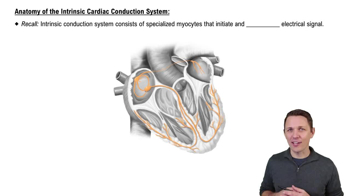
Specialized cardiac cells that spontaneously generate action potentials to establish and conduct the heart's electrical rhythm; SA node is the main pacemaker.
Contractile cardiomyocytes that contract to pump blood in response to electrical signals.
Autorhythmic cells spontaneously depolarize to threshold and fire action potentials.
Contractile cells receive electrical signals via gap junctions and depolarize in response.
Na⁺: depolarization.
Ca²⁺: enters during plateau, triggers contraction.
K⁺: exits causing repolarization.
Calcium is pumped back into sarcoplasmic reticulum during relaxation.
Epinephrine: increases heart rate and contractility.
Calcium-channel blockers: reduce Ca²⁺ entry, lowering heart rate and/or contractility.
Beta blockers: decrease heart rate and contractility by blocking sympathetic stimulation.
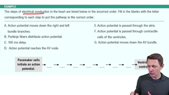
SA node → AV node → AV bundle (His) → right & left bundle branches → Purkinje fibers.
SA node initiates impulse; AV node delays it to allow ventricular filling.
Cardiac output (CO) = blood pumped by one ventricle per minute.
CO = HR × SV.
It determines oxygen and nutrient delivery to tissues.
1. Ventricular filling
2. Atrial systole
3. Isovolumetric contraction
4. Ventricular ejection
5. Isovolumetric relaxation
Systole: contraction phase.
Diastole: relaxation phase.
P wave: atrial depolarization.
QRS complex: ventricular depolarization.
T wave: ventricular repolarization.
Heart sounds are caused by valve closures and resulting vibrations.
LUB (S1): AV valves close.
DUB (S2): semilunar valves close.
Heart murmur is abnormal sound from turbulent flow, often valve-related.
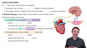
SA node sets rate, modified by autonomic nervous system.
Sympathetic stimulation increases HR.
Parasympathetic (vagus nerve) decreases HR.
Epinephrine also increases HR.
EDV: blood volume in ventricle after filling.
ESV: blood remaining after ejection.
Stroke volume (SV): SV = EDV − ESV.
Factors: preload, contractility, afterload.
Cardiac output (CO) = heart rate (HR) × stroke volume (SV).
If SV increases with constant HR, CO increases.
Exercise increases heart rate to raise cardiac output and oxygen delivery.
Regular aerobic training lowers resting HR due to increased stroke volume.
CHF is when the heart cannot pump effectively, causing blood backup and fluid congestion.
Left-sided failure causes lung edema; right-sided causes systemic tissue edema.
CAD is narrowing/blockage of coronary arteries due to atherosclerosis.
It reduces blood and oxygen to myocardium, causing ischemia, angina, or heart attack.