Skip to main content
General Biology
Il mio corso
Impara
Preparazione agli esami
AI Tutor
Guide di studio
Soluzioni per libri di testo
Flashcard
Esplora
Prova l'app
Il mio corso
Impara
Preparazione agli esami
AI Tutor
Guide di studio
Soluzioni per libri di testo
Flashcard
Esplora
Prova l'app
Indietro
Anatomy and Physiology of the Brain
Puoi toccare per girare la carta.
Cerebrum
Puoi toccare per girare la carta.
👆
Cerebrum
Largest part of the brain
, responsible for sensory recognition, consciousness, reason, planning, and memory.
Avanzamenti del tracciato
I pulsanti di controllo sono stati cambiati in modalità "navigazione".
1/21
Video consigliati
04:26
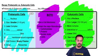
Recap: Prokaryotic vs. Eukaryotic Cells
24476
views
419
rank
05:54
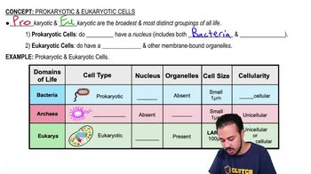
Prokaryotic & Eukaryotic Cells
40684
views
895
rank
4
comments
03:54
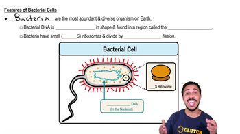
Features of Bacterial Cells
29704
views
524
rank
1
comments
Termini in questo insieme (21)
Nascondere definizioni
Cerebrum
Largest part of the brain
, responsible for sensory recognition, consciousness, reason, planning, and memory.
Gyri
Cerebral ridges
on the surface of the cerebrum that increase surface area.
Sulci
Cerebral clefts or depressions
that separate gyri.
Cerebral lobes
Defined regions of the cerebrum with
specific functions
such as sensory processing and cognition.
Electroencephalography (EEG)
Measurement of brain electrical activity (action potentials) in the cerebral cortex to assess brain function.
Brain Cerebellum
Part of the hindbrain critical for
balance, timing, and coordination
of skeletal muscle contractions.
Diencephalon
Medial brain region controlling behavior and the endocrine system, including the epithalamus, hypothalamus, pituitary gland, and thalamus.
Epithalamus (pineal gland)
Gland that releases
melatonin
and controls circadian and diurnal biological rhythms.
Hypothalamus
Contains nuclei that control autonomic activity, behavior, and pituitary gland function.
Pituitary gland (Hypophysis)
Produces neuroendocrine and endocrine hormones regulating homeostasis and reproduction.
Thalamus
Relay center for sensory information between the cerebrum, brainstem, cerebellum, and spinal cord.
Midbrain
Controls some eye motor movements and is involved in auditory and visual processing.
Pons
Bridge between cerebrum and cerebellum; origin of the trigeminal nerve controlling facial sensation and mastication.
Medulla oblongata
Continuation of spinal cord; controls autonomic functions like heart rate, respiration, and reflexes such as coughing and swallowing.
Ventricular system
Network of brain cavities filled with cerebrospinal fluid (CSF) that cushions the brain and provides nutrients.
Choroid plexus
Capillary network in ventricles covered by ependymal cells that produce cerebrospinal fluid.
Meninges
Three protective connective tissue layers covering brain and spinal cord: dura mater, arachnoid mater, and pia mater.
Dura mater
Tough outer meningeal layer attached to the skull bones.
Arachnoid mater
Transparent membrane between dura and pia mater; creates subarachnoid space where CSF circulates.
Pia mater
Delicate membrane covering the brain and spinal cord surface.
Blood-brain barrier
Cellular barrier controlling molecule transport from blood to brain, allowing only essential substances to maintain neural health.
 IndietroIndietro
 04:26