Skip to main content
General Biology
Il mio corso
Impara
Preparazione agli esami
AI Tutor
Guide di studio
Soluzioni per libri di testo
Flashcard
Esplora
Prova l'app
Il mio corso
Impara
Preparazione agli esami
AI Tutor
Guide di studio
Soluzioni per libri di testo
Flashcard
Esplora
Prova l'app
Indietro
Microscopy and Cell Fractionation in General Biology
Puoi toccare per girare la carta.
What invention allowed cell biologists to investigate cells too small to see with the unaided eye?
Puoi toccare per girare la carta.
👆
What invention allowed cell biologists to investigate cells too small to see with the unaided eye?
The invention of the microscope, first in 1590 and refined in the 1600s, allowed visualization of cells and microorganisms.
Avanzamenti del tracciato
I pulsanti di controllo sono stati cambiati in modalità "navigazione".
1/28
Video consigliati
06:35
Microscopes
24027
views
554
rank
02:52
Types of Electron Microscopes
17200
views
349
rank
1
comments
Termini in questo insieme (28)
Nascondere definizioni
What invention allowed cell biologists to investigate cells too small to see with the unaided eye?
The invention of the microscope, first in 1590 and refined in the 1600s, allowed visualization of cells and microorganisms.
Who first observed cell walls and when?
Robert Hooke first observed cell walls in 1665 by looking at dead cells from oak tree bark.
What type of microscope did Antoni van Leeuwenhoek use to visualize living cells?
Van Leeuwenhoek used early light microscopes with finely crafted lenses to visualize living cells and microorganisms.
What are the three important parameters in microscopy?
Magnification
,
resolution
, and
contrast
.
Define magnification in microscopy.
Magnification is the ratio of an object's image size to its real size.
What is the effective magnification limit of a light microscope?
Light microscopes can magnify effectively up to about 1,000 times the actual size of the specimen.
Define resolution in microscopy.
Resolution is the minimum distance two points can be separated and still be distinguished as separate points.
What is the resolution limit of a standard light microscope?
About 0.2 micrometers (200 nanometers), regardless of magnification.
What does contrast refer to in microscopy?
Contrast is the difference in brightness between the light and dark areas of an image.
How can contrast be enhanced in light microscopy?
By staining or labeling cell components to make them stand out visually.
What is the size range of most cells?
Most cells range between 1 and 100 micrometers in diameter.
What is the size range of viruses compared to cells?
Viruses are smaller, typically between 50 and 100 nanometers.
What is phase-contrast microscopy used for?
It amplifies variations in density within unstained cells to enhance contrast, useful for living, unpigmented cells.
What advantage does differential-interference contrast (Nomarski) microscopy provide?
It exaggerates differences in density to produce almost 3D appearing images.
How does fluorescence microscopy work?
Fluorescent dyes or antibodies label molecules; they absorb UV radiation and emit visible light to reveal specific locations.
What is confocal microscopy?
A laser-based technique that eliminates out-of-focus light to create sharp optical sections and 3D reconstructions.
What is the main difference between light microscopes and electron microscopes?
Electron microscopes use electron beams with shorter wavelengths, allowing much higher resolution than light microscopes.
What is the resolution limit of modern electron microscopes?
Theoretical resolution is about 0.002 nanometers; practical resolution is about 2 nanometers.
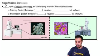
What is the function of a scanning electron microscope (SEM)?
SEM scans the specimen surface with electrons to produce detailed 3D images of surface topography.
What is the function of a transmission electron microscope (TEM)?
TEM transmits electrons through thin specimen sections to study internal cell structures.
Why do electron microscopes require heavy metal staining?
Heavy metals increase electron density in certain structures, enhancing contrast by scattering electrons.
What is a disadvantage of electron microscopy compared to light microscopy?
Electron microscopy preparation kills cells and can introduce artifacts not present in living cells.
What recent advances have improved light microscopy?
Fluorescent markers, confocal microscopy, and super-resolution microscopy have enhanced detail and resolution.
What is cryo-electron microscopy (cryo-EM)?
A TEM technique preserving specimens at very low temperatures to visualize structures in their native environment.
What is cell fractionation?
A technique where broken cells are centrifuged to separate components by size and density into pellets.
How does cell fractionation help study cell function?
It allows isolation of specific organelles in bulk to identify their biochemical functions.
What discovery was made by combining cell fractionation and electron microscopy?
Mitochondria were identified as the sites of cellular respiration.
Why is integrating cytology and biochemistry important?
It correlates cell structure with function by combining microscopic and chemical analyses.
 IndietroIndietro
 06:35
06:35