 Indietro
Microscopy and Cell Fractionation in General Biology
 06:35
06:35
Termini in questo insieme (20)
Magnification is the ratio of an object’s image size to its real size, showing how much larger the image appears compared to the actual specimen.
Resolution is the measure of image clarity, specifically the minimum distance two points can be separated and still be distinguished as separate points.
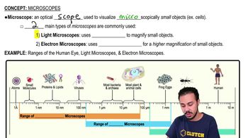
The light microscope cannot resolve details finer than about \(0.2\ \mu m\) (200 nm) due to the wavelength of visible light.
Contrast is the difference in brightness between light and dark areas of an image, enhanced by staining or labeling cell components.
Robert Hooke first observed cell walls in 1665 using a microscope to look at dead cells from oak tree bark.
He crafted lenses that allowed visualization of living cells and microorganisms, called "very little animalcules," in 1674.
Visible light passes through the specimen and glass lenses, which refract the light to magnify the image projected to the eye or camera.
Most cells range between 1 and 100 micrometers (\(\mu m\)) in diameter.
EM uses a beam of electrons instead of light to achieve much higher resolution due to electrons’ shorter wavelengths.
Electron microscopes can theoretically resolve structures as small as about \(0.002\ nm\), but practically about 2 nm.
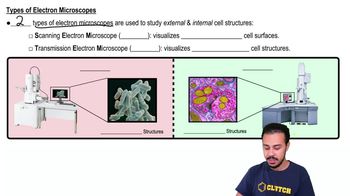
SEM provides detailed 3D images of the surface topography of specimens by scanning with an electron beam.
TEM transmits electrons through a thin specimen section to study internal cell structures, while SEM scans surfaces.
Heavy metals increase electron density in certain structures, enhancing contrast by scattering electrons more in denser regions.
Electron microscopy requires specimen preparation that kills cells and may introduce artifacts, unlike light microscopy which can study living cells.
Fluorescent markers, confocal microscopy, and super-resolution techniques have improved detail and resolution beyond traditional limits.
Cryo-EM preserves specimens at very low temperatures without preservatives, allowing visualization of structures in their natural cellular environment.
Cell fractionation is a technique where broken cells are centrifuged to separate components by size and density into pellets for study.
Spinning broken cells at increasing speeds causes larger components to pellet first, followed by smaller components in subsequent spins.
Biochemical tests on fractions with mitochondria showed enzymes for cellular respiration, linking mitochondria to energy production.
Microscopy allows visualization of cell structures, essential for understanding cell function and integrating with biochemical data.