Endocrine signals travel through the bloodstream to distant targets. Paracrine signals act locally on nearby cells. Autocrine signals act on the same cell that produces them. Juxtacrine signals require direct cell contact.
Definition of ligand and receptor
A ligand is a chemical messenger that binds specifically to a receptor on or inside a target cell to initiate a response.
Signal transduction
The process by which ligand binding to a receptor triggers a cascade of intracellular events, often involving second messengers, leading to cellular responses or changes in gene expression.
Receptor-ligand binding characteristics
Binding is highly specific and reversible, resembling enzyme-substrate interactions. Saturation occurs when most receptors are occupied, described by receptor affinity.
Small amounts of ligand can produce large cellular responses because each step in the signaling cascade activates multiple downstream molecules.
Hydrophilic vs hydrophobic ligands
Hydrophilic ligands bind transmembrane receptors; hydrophobic ligands cross membranes and bind intracellular receptors, often regulating transcription.
G protein-coupled receptors (GPCRs)
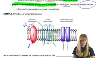
Receptors with seven transmembrane helices that activate heterotrimeric G proteins upon ligand binding, initiating intracellular signaling.
G protein activation cycle
Inactive G protein bound to GDP associates with receptor; ligand binding causes GDP release, GTP binding, and dissociation of Gα and Gβγ subunits to activate effectors.
Second messengers in G protein signaling
Common second messengers include cyclic AMP (cAMP) and calcium ions (Ca2+), which relay and amplify signals inside the cell.
cAMP production and function
cAMP is synthesized from ATP by adenylyl cyclase activated by GTP-bound Gα. It activates protein kinase A (PKA), which phosphorylates target proteins.
Phospholipase C pathway
Activated by Gq proteins, phospholipase C cleaves PIP2 into IP3 and DAG. IP3 releases Ca2+ from ER; DAG and Ca2+ activate protein kinase C (PKC).
Calcium signaling
Ca2+ is tightly regulated and released from ER stores via IP3 receptors. It binds calmodulin and other proteins to regulate diverse cellular processes.
Receptor tyrosine kinases (RTKs)
Single-pass transmembrane receptors with intrinsic kinase activity that dimerize and autophosphorylate upon ligand binding, triggering downstream signaling.
Ras-MAP kinase pathway
Activated RTKs recruit adaptor proteins to activate Ras, a small GTPase, which triggers a kinase cascade (Raf → MEK → MAPK) leading to gene expression changes.
Dominant negative and constitutively active receptor mutations
Dominant negative mutations interfere with normal receptor function; constitutively active mutations cause receptors to signal without ligand, often linked to disease.
Cytoskeletal elements in eukaryotic cells
Three main types: microtubules (25 nm, tubulin), microfilaments (7 nm, actin), and intermediate filaments (8–12 nm, variable proteins).
Microtubule structure and polarity
Microtubules are hollow cylinders of 13 protofilaments made of αβ-tubulin heterodimers, with a plus end (fast-growing) and a minus end (slow-growing).
Dynamic instability of microtubules
Microtubules alternate between growth and shrinkage phases controlled by GTP hydrolysis on β-tubulin, with 'catastrophe' and 'rescue' events.
Microtubule-organizing centers (MTOCs)
Sites like centrosomes nucleate and anchor microtubules, with γ-tubulin ring complexes initiating microtubule assembly.
Microfilament (actin) structure and dynamics
Actin monomers (G-actin) polymerize into polarized filaments (F-actin) with ATP-actin at the plus end; assembly regulated by actin-binding proteins.
Actin-binding proteins functions
Regulate nucleation, elongation, severing, capping, bundling, and crosslinking of actin filaments to form diverse cellular structures.
Rho family GTPases in actin regulation
Rho, Rac, and Cdc42 regulate actin cytoskeleton remodeling: Rho promotes stress fibers, Rac lamellipodia, and Cdc42 filopodia formation.
Intermediate filaments
Stable cytoskeletal components providing mechanical strength; tissue-specific proteins like keratins and lamins assemble into coiled-coil dimers and filaments.
Motor proteins and cellular motility
Kinesins and dyneins move cargo along microtubules; myosins move along actin filaments, enabling intracellular transport, muscle contraction, and cell movement.
Muscle contraction mechanism
Myosin II heads bind actin filaments and use ATP hydrolysis to generate force, sliding thin filaments past thick filaments in sarcomeres.
Regulation of muscle contraction by calcium
Calcium binds troponin, causing tropomyosin to expose myosin-binding sites on actin, enabling cross-bridge cycling and contraction.
Cell-cell junction types
Adhesive junctions (adherens, desmosomes), tight junctions (seal cells), and gap junctions (allow small molecule passage) maintain tissue integrity.
Extracellular matrix (ECM) components
Includes structural proteins (collagen, elastin), proteoglycans (hydrated matrix), and adhesive glycoproteins (fibronectin, laminin) linking cells to ECM.
Integrins
Transmembrane receptors that bind ECM proteins and connect to the cytoskeleton, mediating adhesion and signaling.
 Indietro
 07:35
