Staining provides contrast to promote visibility of bacteria under the microscope.
Three key features of a microscope for viewing bacteria
Magnification, resolution, and contrast.
Difference between simple and differential staining
Simple staining uses one stain to show shape and arrangement; differential staining uses multiple stains to identify bacteria by cell wall characteristics.
Four essential steps in bacterial staining procedure
Use a sterile inoculation loop to place a loopful of broth on a clean slide and spread thinly over an area the size of a dime.
How to prepare a smear from solid culture
Place a drop of water or saline on slide, transfer bacteria from colony with loop, mix in circular motion to size of a nickel.
Purpose of heat fixation
Kills bacteria and affixes the smear to the slide so it does not wash off during staining.
Components of a chromogen in staining
Chromogen has a chromophore (color generating) and an auxochrome (charged component that binds cells).
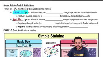
Difference between basic and acidic dyes
Basic dyes are positively charged and stain negatively charged bacterial surfaces; acidic dyes are negatively charged and stain the background (negative staining).
What is positive staining?
Staining where the microbe surface is stained due to attraction of basic dyes to negatively charged cells.
What is negative staining?
Staining where the background is stained by acidic dyes, leaving cells unstained and visible.
Common bacterial shapes observed in simple staining
Coccus (round), Bacillus (rod-shaped), Spiral forms including vibrio, spirillum, and spirochete.
Description of coccus bacteria
Spherical or round-shaped bacteria.
Description of bacillus bacteria
Rod-shaped bacteria.
Types of spiral bacteria
Vibrio (comma-shaped), spirillum (curved or spiral), and spirochete (corkscrew-shaped).
 Indietro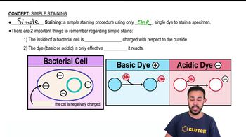
 03:23
03:23