 Indietro
The Integumentary System: Structure, Function, and Clinical Aspects
Guida di studio - Note intelligenti
 Appunti personalizzati basati sui tuoi materiali, ampliati con definizioni chiave, esempi e contesto.
The Integumentary System
Overview and Major Regions
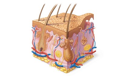
The integumentary system, commonly referred to as the skin, is the body's largest organ and serves as a protective covering. It consists of three major regions: the epidermis (superficial), dermis (middle), and hypodermis (deepest, also called subcutaneous layer). The hypodermis is primarily composed of adipose tissue and is not technically part of the skin.
Structure of the Skin
The skin is organized into distinct layers, each with specialized functions and cell types:
Epidermis: The outermost layer, composed of keratinized stratified squamous epithelium. It is avascular and contains four main cell types: keratinocytes, melanocytes, dendritic cells, and tactile (Merkel) cells.
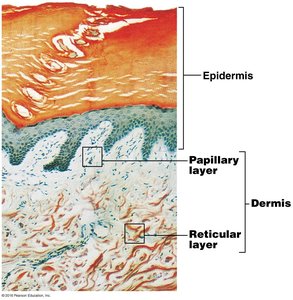
Dermis: A strong, flexible connective tissue layer containing fibroblasts, macrophages, mast cells, and white blood cells. It is divided into two layers: the papillary layer (areolar connective tissue) and the reticular layer (dense irregular connective tissue).
Hypodermis: A subcutaneous layer mainly composed of adipose tissue, providing insulation and energy storage.
Epidermal Layers and Cell Types
Layers of the Epidermis
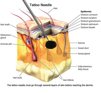
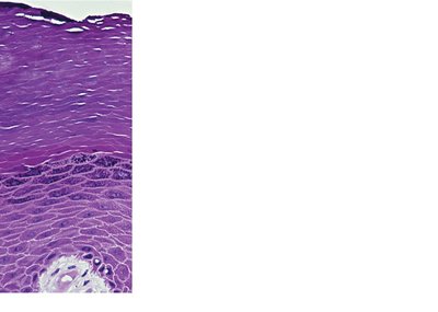
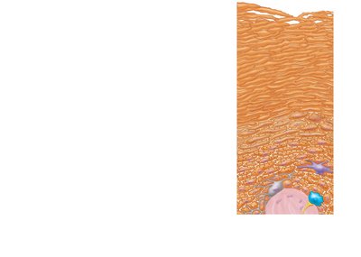
The epidermis is organized into four (sometimes five) distinct layers, from superficial to deep:
Stratum corneum: 20–30 layers of dead, keratin-filled cells providing protection.
Stratum granulosum: 3–5 layers of flattened cells where keratinization begins and organelles deteriorate.
Stratum spinosum: Several layers of keratinocytes unified by desmosomes; contains dendritic cells.
Stratum basale: A single row of actively mitotic stem cells; contains melanocytes and tactile cells.
Cell Types of the Epidermis
Keratinocytes: Most abundant; produce keratin, a protein that provides strength and waterproofing.
Melanocytes: Produce melanin pigment, which protects against UV radiation.
Dendritic (Langerhans) cells: Macrophages that help activate the immune system.
Tactile (Merkel) cells: Sensory receptors for touch.
Dermis: Structure and Function
Papillary Layer
The papillary layer is the thinner, superficial part of the dermis, composed of areolar connective tissue. It contains dermal papillae, which house capillary loops, Meissner’s corpuscles (touch receptors), and free nerve endings (pain receptors).
Reticular Layer
The reticular layer is the deeper, thicker part of the dermis, composed of dense irregular connective tissue. It provides strength, resiliency, and elasticity due to its collagen and elastic fibers. Blood vessels are found between this layer and the hypodermis.
Stretch Marks
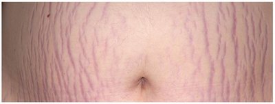
Stretch marks (striae) result from tearing of the dermis, often due to rapid growth or weight changes.
Skin Color
Pigments Contributing to Skin Color
Melanin: Ranges from yellow to reddish-brown to black; produced by melanocytes in the stratum basale. Responsible for dark skin colors, freckles, and pigmented moles.
Carotene: Yellow to orange pigment, most obvious in the palms and soles.
Hemoglobin: Provides the pinkish hue of skin, especially in fair-skinned individuals.
Appendages of the Skin
Sweat Glands
Sweat glands are derived from the epidermis and are classified as:
Eccrine sweat glands: Abundant on palms, soles, and forehead; produce watery sweat for thermoregulation. Ducts connect directly to pores.
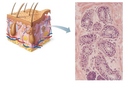
Apocrine sweat glands: Confined to axillary and anogenital areas; secrete sweat plus fatty substances and proteins. Ducts connect to hair follicles. Specialized apocrine glands include ceruminous (ear wax) and mammary glands.
Sebaceous (Oil) Glands
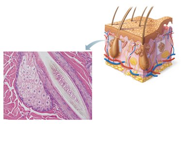
Widely distributed, most develop from hair follicles, and become active at puberty. They secrete sebum, an oily substance that is bactericidal and softens hair and skin.
Hair (Pili)
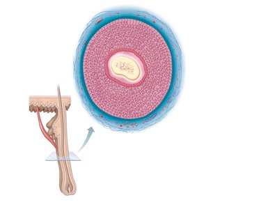
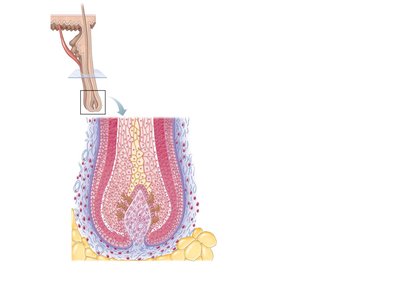
Hair consists of dead, keratinized cells and contains hard keratin. Hair pigments are produced by melanins. Gray/white hair results from decreased melanin production and increased air bubbles in the shaft. The hair follicle is composed of several layers, including the cuticle, cortex, and medulla.
Arrector Pili Muscle
The arrector pili is a smooth muscle attached to the hair follicle, responsible for causing "goose bumps" by contracting and pulling the hair upright.
Nails
Nails are scalelike modifications of the epidermis, composed of hard keratin. They are used for diagnosis of certain conditions (e.g., malnutrition, iron deficiency, fungal infection, respiratory or thyroid disorders).
Functions of the Integumentary System
Protection
The skin provides three types of barriers:
Chemical barriers: Acid mantle and defensins slow or kill bacteria; melanin protects against UV damage.
Physical/mechanical barriers: Keratin and glycolipids block most water and water-soluble substances; limited penetration by certain chemicals.
Biological barriers: Dendritic cells, macrophages, and DNA contribute to immune defense.
Body Temperature Regulation
The skin regulates body temperature through perspiration and dilation of dermal blood vessels.
Cutaneous Sensations
The skin contains receptors for temperature, touch, and pain.
Metabolic Functions
The skin synthesizes vitamin D precursor and collagenase, and chemically converts carcinogens and some hormones.
Blood Reservoir and Excretion
The skin can hold up to 5% of the body's blood volume and excretes nitrogenous wastes and salt in sweat.
Burns and Clinical Aspects
Burns
Burns are caused by heat, electricity, radiation, or chemicals, resulting in tissue damage, denatured proteins, and cell death. The immediate threat is loss of fluids and infection, which can lead to dehydration, electrolyte imbalance, renal shutdown, and circulatory shock.
Rule of Nines
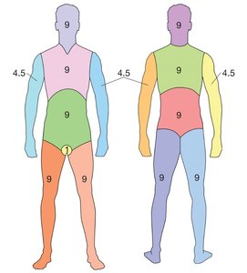
The rule of nines is used to estimate the extent of burns on the body, dividing it into regions that represent 9% (or multiples) of total body surface area.
Types of Burns
First-degree: Epidermal damage only; redness, edema, pain; heals in 2–3 days.
Second-degree: Epidermal and upper dermal damage; blisters; heals in 3–4 weeks.
Third-degree: Entire thickness of skin damaged; gray-white, cherry red, or black; no initial pain (nerve endings destroyed); skin grafting usually necessary.
Severity and Complications
Burns are critical if >25% of the body has second-degree burns, >10% has third-degree burns, or if face, hands, or feet bear third-degree burns. Infection (sepsis) is a leading cause of death.
Diseases of the Skin
Common Skin Diseases
Impetigo: Bacterial infection (staph or strep), highly contagious.
Fifth disease: Viral infection (parvovirus), characterized by "slapped cheek" appearance.
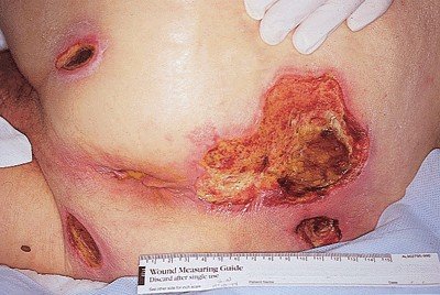
Bedsores (Decubitus ulcers): Caused by prolonged pressure, leading to tissue necrosis.
Viral Skin Diseases
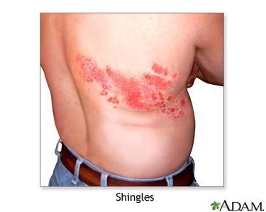
Shingles: Reactivation of the chickenpox virus, dormant in nerves; causes painful rash.
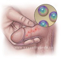
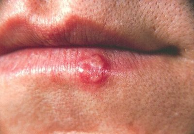
Herpes Simplex: Virus, contagious, dormant in nerves; Type 1 (oral, cold sores), Type 2 (genital).
Skin Cancer
Types of Skin Cancer
Basal cell carcinoma: Least malignant, most common; arises from stratum basale cells.
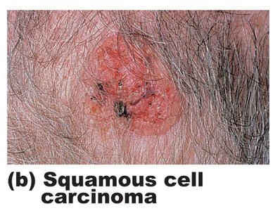
Squamous cell carcinoma: Second most common; involves keratinocytes of stratum spinosum; good prognosis if treated early.
Melanoma: Cancer of melanocytes; highly metastatic and resistant to chemotherapy; treated by wide surgical excision and immunotherapy.
Additional info: The notes above expand on the original content by providing definitions, examples, and clinical context for each topic, ensuring completeness and academic quality for exam preparation.
