Ch. 17 The Special Senses
Martini - Fundamentals of Anatomy & Physiology 12th Edition
Martini, Nath, Bartholomew12th EditionFundamentals of Anatomy & PhysiologyISBN: 9780137854011Non è quello che usi tu?Cambia libro di testo
Capitolo 17, Problema 10

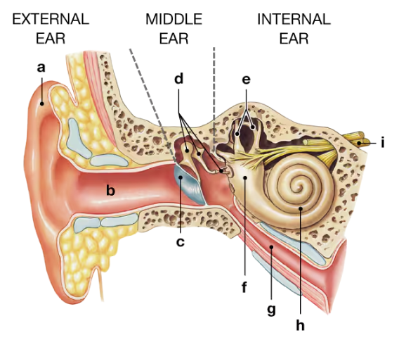
Identify the structures of the external, middle, and internal ear in the following figure.
Diagram of the external, middle, and internal ear with labeled anatomical structures a through i.
a. ___
b. ___
c. ___
d. ___
e. ___
f. ___
g. ___
h. ___
i. ___

Guida verificata passo dopo passo
1
Step 1: Understand the three main regions of the ear: the external ear, middle ear, and internal ear. The external ear includes structures that collect sound waves, the middle ear contains ossicles that transmit vibrations, and the internal ear houses sensory organs for hearing and balance.
Step 2: Identify the external ear structures, which typically include the auricle (pinna) and the external auditory canal. These are the parts visible from outside and lead sound waves inward.
Step 3: Locate the middle ear components, which are found within the tympanic cavity. These include the tympanic membrane (eardrum) and the three auditory ossicles: malleus, incus, and stapes. These bones amplify and transmit sound vibrations to the inner ear.
Step 4: Recognize the internal ear structures, which consist of the cochlea (responsible for hearing), vestibule, and semicircular canals (both involved in balance). These are housed within the bony labyrinth of the temporal bone.
Step 5: Match each labeled part (a through i) in the figure to these categories by comparing their location and shape to the known anatomy of the ear regions, assigning each label to either external, middle, or internal ear structures accordingly.

Risposta video verificata per un problema simile:

Questa soluzione video è stata consigliata dai nostri tutor come utile per risolvere questo problema.

Concetti chiave

Ecco i concetti essenziali che devi comprendere per rispondere correttamente alla domanda.

Anatomy of the External Ear

The external ear includes the auricle (pinna) and the external auditory canal. It collects sound waves and directs them toward the tympanic membrane (eardrum). Understanding its structures helps identify parts visible or accessible from outside the head.
Video consigliato:
3:44
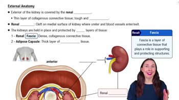
External Anatomy

Anatomy of the Middle Ear

The middle ear is an air-filled cavity containing the tympanic membrane and ossicles (malleus, incus, stapes). These bones transmit sound vibrations from the eardrum to the inner ear. Recognizing these components is essential for identifying the middle ear structures.
Video consigliato:
4:14
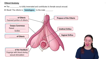
Clitoral Anatomy

Anatomy of the Internal Ear

The internal ear consists of the cochlea, vestibule, and semicircular canals, responsible for hearing and balance. It converts mechanical vibrations into nerve impulses. Knowing these parts aids in distinguishing the internal ear structures in diagrams.
Video consigliato:
6:14
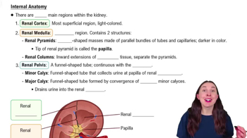
Internal Anatomy