Ch. 11 - Gene Mutation, DNA Repair, and Homologous Recombination
Sanders - Genetic Analysis: An Integrated Approach 3rd Edition
Sanders3rd EditionGenetic Analysis: An Integrated ApproachISBN: 9780135564172Non è quello che usi tu?Cambia libro di testo
Capitolo 11, Problema 31

Experiments by Charles Yanofsky in the 1950s and 1960s helped characterize the nature of tryptophan synthesis in E. coli. In one of Yanofsky's experiments, he identified glycine (Gly) as the wild-type amino acid in position 211 of tryptophan synthetase, the product of the trpA gene. He identified two independent missense mutants with defective tryptophan synthetase at these positions that resulted from base-pair substitutions. One mutant encoded arginine (Arg) and another encoded glutamic acid (Glu). At position 235, wild-type tryptophan synthetase contains serine (Ser) but a base-pair substitution mutant encodes leucine (Leu). At position 243, the wild-type polypeptide contains glutamine and a base-pair substitution mutant encodes a stop codon. Identify the most likely wild-type codons for positions 211, 235, and 243. Justify your answer in each case.

Guida verificata passo dopo passo
1
Step 1: Understand the problem. The question asks us to identify the most likely wild-type codons for positions 211, 235, and 243 in the trpA gene of E. coli based on the amino acids encoded in the wild-type and mutant forms. This involves analyzing the genetic code and determining which codons correspond to the specified amino acids.
Step 2: Analyze position 211. The wild-type amino acid is glycine (Gly), and the mutants encode arginine (Arg) and glutamic acid (Glu). Glycine is encoded by codons GGU, GGC, GGA, and GGG. Arginine is encoded by codons CGU, CGC, CGA, CGG, AGA, and AGG. Glutamic acid is encoded by codons GAA and GAG. To determine the most likely wild-type codon, consider which glycine codon could mutate to encode arginine or glutamic acid through a single base-pair substitution.
Step 3: Analyze position 235. The wild-type amino acid is serine (Ser), and the mutant encodes leucine (Leu). Serine is encoded by codons UCU, UCC, UCA, UCG, AGU, and AGC. Leucine is encoded by codons UUA, UUG, CUU, CUC, CUA, and CUG. To determine the most likely wild-type codon, consider which serine codon could mutate to encode leucine through a single base-pair substitution.
Step 4: Analyze position 243. The wild-type amino acid is glutamine (Gln), and the mutant encodes a stop codon. Glutamine is encoded by codons CAA and CAG. Stop codons are UAA, UAG, and UGA. To determine the most likely wild-type codon, consider which glutamine codon could mutate to a stop codon through a single base-pair substitution.
Step 5: Justify the answers. For each position, the most likely wild-type codon is the one that can mutate to the observed mutant codons with minimal changes in the nucleotide sequence. Use the genetic code table to verify the possible single base-pair substitutions that result in the observed amino acid changes or stop codon.

Risposta video verificata per un problema simile:

Questa soluzione video è stata consigliata dai nostri tutor come utile per risolvere questo problema.
Durata del video:
2m

Concetti chiave

Ecco i concetti essenziali che devi comprendere per rispondere correttamente alla domanda.

Codons and Amino Acids

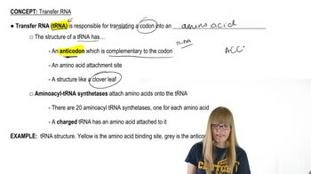
Codons are sequences of three nucleotides in mRNA that correspond to specific amino acids during protein synthesis. Each amino acid is encoded by one or more codons, and understanding the genetic code is essential for determining which codons correspond to the wild-type amino acids in the context of mutations. For example, the codon for glycine (Gly) is GGC, while serine (Ser) is encoded by UCU, UCC, UCA, or UCG.
Video consigliato:

Missense Mutations

Missense mutations occur when a single nucleotide change results in the substitution of one amino acid for another in a protein. This type of mutation can affect protein function depending on the properties of the substituted amino acid. In Yanofsky's experiments, the identification of missense mutants at specific positions in tryptophan synthetase illustrates how base-pair substitutions can lead to different amino acids, impacting the enzyme's activity.
Video consigliato:
Percorso guidato
09:49
Point Mutations

Base-Pair Substitution

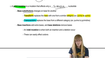
Base-pair substitutions are a type of mutation where one nucleotide in the DNA sequence is replaced by another. This can lead to various outcomes, including silent mutations (no change in amino acid), missense mutations (change in one amino acid), or nonsense mutations (introduction of a stop codon). In the context of the question, understanding how these substitutions affect the amino acid sequence of tryptophan synthetase is crucial for identifying the wild-type codons.
Video consigliato:
Percorso guidato
03:49
Base Distortions
Pratica correlata
Domanda del libro di testo

Alkaptonuria is a human autosomal recessive disorder caused by mutation of the HAO gene that encodes the enzyme homogentisic acid oxidase. A map of the HAO gene region reveals four BamHI restriction sites (B1 to B4) in the wild-type allele and three BamHI restriction sites in the mutant allele. BamHI utilizes the restriction sequence 5′-GGATCC-3′. The BamHI restriction sequence identified as B3 is altered to 5′-GGAACC-3′ in the mutant allele. The mutation results in a Ser-to-Thr missense mutation. Restriction maps of the two alleles are shown below, and the binding sites of two molecular probes (probe A and probe B) are identified.

DNA samples taken from a mother (M), father (F), and two children (C1 and C2) are analyzed by Southern blotting of BamHI-digested DNA. The gel electrophoresis results are illustrated.

Explain how the DNA sequence change results in a Ser-to-Thr missense mutation.

535
views
Domanda del libro di testo

A fragment of a wild-type polypeptide is sequenced for seven amino acids. The same polypeptide region is sequenced in four mutants.

Identify the mutation that produces each mutant polypeptide.

416
views
Domanda del libro di testo

A fragment of a wild-type polypeptide is sequenced for seven amino acids. The same polypeptide region is sequenced in four mutants.

Use the available information to characterize each mutant.

524
views
Domanda del libro di testo

Alkaptonuria is a human autosomal recessive disorder caused by mutation of the HAO gene that encodes the enzyme homogentisic acid oxidase. A map of the HAO gene region reveals four BamHI restriction sites (B1 to B4) in the wild-type allele and three BamHI restriction sites in the mutant allele. BamHI utilizes the restriction sequence 5′-GGATCC-3′. The BamHI restriction sequence identified as B3 is altered to 5′-GGAACC-3′ in the mutant allele. The mutation results in a Ser-to-Thr missense mutation. Restriction maps of the two alleles are shown below, and the binding sites of two molecular probes (probe A and probe B) are identified.

DNA samples taken from a mother (M), father (F), and two children (C1 and C2) are analyzed by Southern blotting of BamHI-digested DNA. The gel electrophoresis results are illustrated.

In a separate figure, draw the gel electrophoresis band patterns for all the genotypes that could be found in children of this couple.

423
views
Domanda del libro di testo

A fragment of a wild-type polypeptide is sequenced for seven amino acids. The same polypeptide region is sequenced in four mutants.

Determine the wild-type mRNA sequence.

579
views
Domanda del libro di testo

Alkaptonuria is a human autosomal recessive disorder caused by mutation of the HAO gene that encodes the enzyme homogentisic acid oxidase. A map of the HAO gene region reveals four BamHI restriction sites (B1 to B4) in the wild-type allele and three BamHI restriction sites in the mutant allele. BamHI utilizes the restriction sequence 5′-GGATCC-3′. The BamHI restriction sequence identified as B3 is altered to 5′-GGAACC-3′ in the mutant allele. The mutation results in a Ser-to-Thr missense mutation. Restriction maps of the two alleles are shown below, and the binding sites of two molecular probes (probe A and probe B) are identified.

DNA samples taken from a mother (M), father (F), and two children (C1 and C2) are analyzed by Southern blotting of BamHI-digested DNA. The gel electrophoresis results are illustrated.

Using A to represent the wild-type allele and a for the mutant allele, identify the genotype of each family member. Identify any family member who is alkaptonuric.

569
views