 Indietro
Bacterial Cell Wall Structures and Gram Staining
 01:58
01:58
Termini in questo insieme (27)
Yes, Gram-positive bacteria have a cell wall.
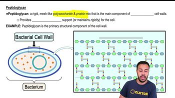
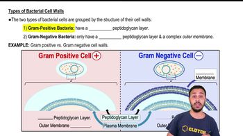
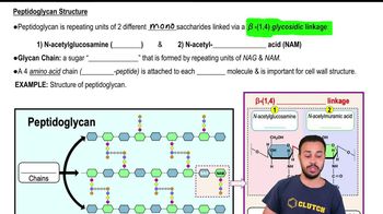
Gram-positive bacteria have a thick peptidoglycan layer.
No, Gram-positive bacteria lack an outer membrane.
Yes, Gram-positive bacteria contain teichoic acids in their cell walls.
Gram-positive bacteria stain purple after Gram staining.
Yes, Gram-negative bacteria have a cell wall.
Gram-negative bacteria have a thin peptidoglycan layer.
Yes, Gram-negative bacteria have an outer membrane.
Yes, Gram-negative bacteria have LPS in their outer membrane.
LPS is also called endotoxin.
Gram-negative bacteria stain pink/red after Gram staining.
Gram-negative bacteria are generally more resistant.
Because of their outer membrane which acts as a barrier.
Mycolic acid is present in acid-fast bacterial cell walls.
They have a waxy cell wall that resists staining.
Their waxy cell wall provides protection against many treatments.
No, Mycoplasma lack a cell wall.
Mycoplasma membranes contain sterols.
Because it has no cell wall, penicillin is ineffective.
No, L-form bacteria lack a normal cell wall.
Yes, L-form bacteria are flexible due to lack of a rigid cell wall.
Gram stain separates bacteria based on their cell wall structure.
The first dye is crystal violet.
Iodine acts as the mordant to fix the dye.
Alcohol/decolorizer removes the crystal violet from Gram-negative cells.
Safranin is used as the counterstain to color Gram-negative cells pink/red.
They help identify bacteria and guide appropriate treatment.