 Indietro
Microscopy in Microbiology
 06:13
06:13
Termini in questo insieme (28)
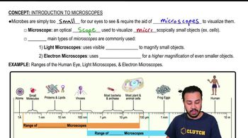
Magnification is the ability of a microscope to produce an image of an object larger than its actual size, allowing more details to be seen than with the unaided eye.
Total magnification = Objective lens magnification × Eyepiece lens magnification.
The resolution limit is approximately 0.2 micrometers (200 nm), known as the diffraction barrier.
It limits the ability to distinguish two objects closer than about half the wavelength of light used, roughly 200 nm for visible light.
The refractive index measures how much a substance slows down light, affecting how light bends when passing through lenses.
Shorter focal length means stronger lens and higher magnification.
Bright-field, dark-field, phase-contrast, fluorescence, and confocal microscopes.
Light passes through the specimen, producing a dark image against a bright background.
NA is a property of the objective lens that determines how much light enters and the resolution; higher NA means better resolution.
d = \(\frac{\lambda}{2NA}\), where λ is wavelength and NA is numerical aperture.
Shorter wavelengths provide better resolution because they allow distinguishing smaller details.
Oil immersion increases numerical aperture and resolution by reducing light refraction between the slide and lens.
Dark-field, phase-contrast, and differential interference contrast (DIC) microscopes.
It forms an image by light reflected or refracted by the specimen, producing a bright image on a dark background.
It enhances contrast by combining deviated and undeviated light, allowing visualization of live cells and internal structures without staining.
It uses fluorochromes to stain specific cell components, which emit visible light when excited, aiding identification of pathogens and proteins.
A laser-based fluorescence technique that creates sharp 3D images by eliminating out-of-focus light.
Electron microscopes use electron beams with much shorter wavelengths, providing higher resolution than light microscopes.
Transmission Electron Microscope (TEM) and Scanning Electron Microscope (SEM).
TEM passes electrons through thin specimens to form 2D images; SEM scans the surface to produce 3D images of surface features.
A technique that rapidly freezes samples to preserve structure and combines multiple TEM images to create 3D molecular images.
It uses a sharp probe to measure surface features at atomic or molecular scale, including scanning tunneling and atomic force microscopes.
Fixation preserves cell structures by inactivating enzymes and toughening cells, using heat or chemical methods depending on the specimen.
Basic dyes have positive charges and bind to negatively charged cell parts; acidic dyes have negative charges and bind to positively charged structures.
A differential stain that divides bacteria into Gram-positive and Gram-negative based on cell wall structure.
It detects bacteria with waxy cell walls like Mycobacterium species, which resist decolorization by acid-alcohol.
Visualizing capsules by staining the background, leaving capsules colorless and visible as halos around cells.
Flagella staining uses mordants to thicken flagella, making them visible under the microscope.