 뒤로
Anatomy & Physiology: Articulations & Body Movements
 04:06

이 집합의 용어 (21)
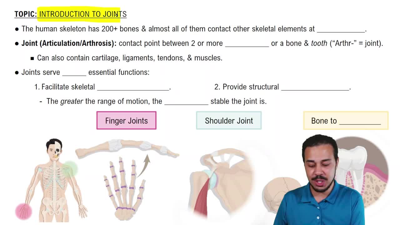
Hold bones together and allow flexibility for body movement.
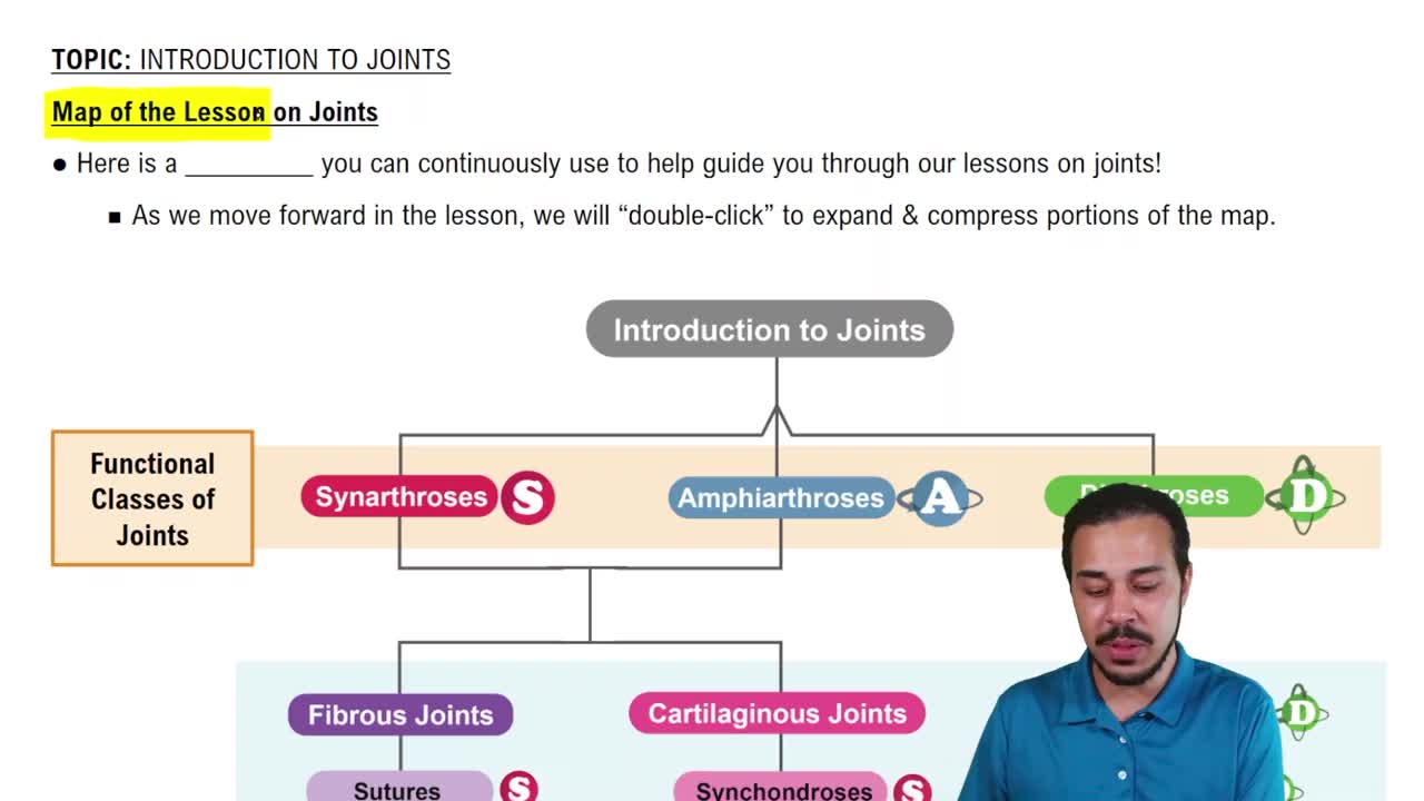
Based on presence of connective fiber, cartilage, or joint cavity: Fibrous, Cartilaginous, Synovial.
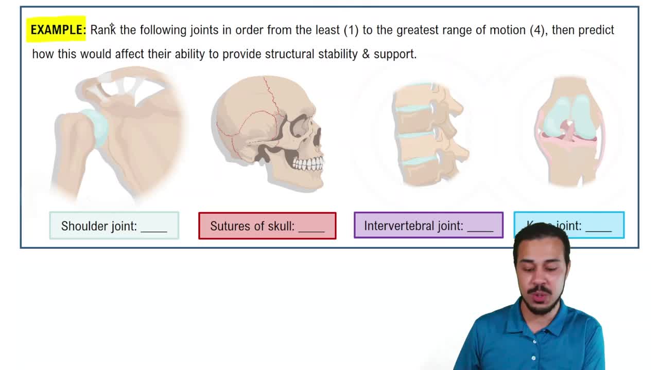
Based on movement: Synarthroses (immovable), Amphiarthroses (slightly movable), Diarthroses (movable).
Connect adjoining bones, lack joint cavity, mostly synarthrotic (immovable).
Sutures (skull bones), Syndesmoses (ligament connections), Gomphosis (tooth in socket).
Bone ends connected by cartilage, lack joint cavity, mostly amphiarthrotic (slightly movable).
Symphyses (fibrocartilage discs, e.g. intervertebral), Synchondroses (hyaline cartilage, e.g. epiphyseal plate).
Articulating bones separated by joint cavity with synovial fluid, all are movable joints.
Articular cartilage, articular capsule (fibrous layer + synovial membrane), synovial fluid, ligaments, nerves, blood vessels.
Composed of fibrocartilage, they reduce wear and tear on joint surfaces; not always present.
Bursa is a synovial fluid-filled sac where tendons cross bones; tendon sheath is an elongated bursa around a tendon.
Uniaxial (one axis), Biaxial (two axes), Multiaxial (three axes).
Plane (gliding), Hinge (flexion/extension), Pivot (rotation), Condylar and Saddle (biaxial), Ball-and-Socket (multiaxial).
Origin is the immovable or less movable attachment; insertion is the movable attachment.
Flexion, Extension, Abduction, Adduction, Rotation, Circumduction.
Large ball-and-socket joint formed by femur head and acetabulum; reinforced by strong ligaments including ligamentum teres.
Three joints: Tibiofemoral (hinge), Femoropatellar, and lateral/medial joints; largest and most complex joint.
Patellar ligament, Medial and Lateral Patellar Retinacula, Fibular and Tibial Collateral, Oblique and Arcuate Popliteal, ACL, and PCL.
ACL prevents tibia sliding forward and provides rotational stability; PCL is stronger, prevents backward tibia movement.
Ball-and-socket joint with greatest range of motion; head of humerus articulates with shallow glenoid cavity; stabilized by rotator cuff muscles.
Modified hinge joint where condylar process of mandible articulates with temporal bone's mandibular fossa, located anterior to ear.