뒤로

Anatomy & Physiology: Blood Vessels and Heart Study Guide

컨트롤 버튼이 '내비게이션' 모드로 변경되었습니다.
1/41
  • Three types of blood vessels

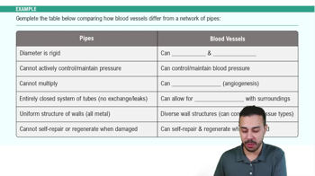
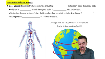
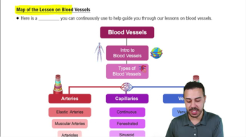
    Arteries carry blood away from the heart, veins carry blood toward the heart, and capillaries exchange substances between blood and tissues.
  • Definition of artery and vein

    Arteries and veins are defined by direction of blood flow, not oxygen content.
  • Three tunics of blood vessels

    Tunica interna: innermost endothelium; Tunica media: smooth muscle controlling diameter; Tunica externa: connective tissue anchoring vessel.
  • Capillary wall structure

    Capillaries have only the tunica interna, one cell layer thick, allowing exchange.
  • Vasa vasorum function

    Tiny vessels in the tunica externa that supply nutrients to large artery walls too thick for diffusion.
  • Three groups of arteries

    Elastic arteries: large, conduct blood; Muscular arteries: distribute blood, active in vasoconstriction; Arterioles: smallest, control flow into capillaries.
  • Role of elastic arteries in blood flow

    Elastic arteries stretch during systole and recoil during diastole, converting pulsatile flow into steady capillary flow.
  • Types of capillaries

    Continuous: tight junctions, skin/muscle; Fenestrated: pores, kidney/intestine; Sinusoidal: large gaps, marrow/liver.
  • Capillary bed components

    Includes vascular shunt (direct arteriole-venule channel) and precapillary sphincters controlling blood flow into true capillaries.
  • Vein characteristics

    Veins have thinner tunica media, larger lumen, contain valves, and act as blood reservoirs holding ~60% of blood.
  • Mechanisms aiding venous return

    Venous valves ensure one-way flow; skeletal muscle pump squeezes veins; respiratory pump changes thoracic pressure.
  • Vascular anastomosis

    Connections between vessels providing alternative blood flow routes to prevent tissue ischemia.
  • Pulmonary circulation pathway

    Right ventricle → pulmonary trunk → pulmonary arteries → lungs → pulmonary veins → left atrium.
  • Systemic circulation pathway

    Left ventricle → aorta → arteries → tissues → venae cavae → right atrium.
  • Blood flow formula

    Blood flow = pressure gradient (ΔP) ÷ resistance (R); flow is driven by pressure difference, not absolute pressure.
  • Three factors affecting resistance

    Vessel length (constant), blood viscosity (slow change), and vessel radius (rapid, major effect).
  • Relationship between vessel radius and resistance

    Resistance ∝ 1 ÷ radius⁴; doubling radius decreases resistance 16-fold.
  • Mean arterial pressure (MAP) formula

    MAP = diastolic pressure + ⅓ pulse pressure; also MAP = cardiac output × total peripheral resistance.
  • Baroreceptor reflex function

    Baroreceptors in aortic arch and carotid sinus detect arterial pressure changes and adjust heart rate and vessel diameter.
  • Effect of low blood pressure on baroreceptors

    Less stretch → decreased firing → increased sympathetic activity → vasoconstriction, increased heart rate and contractility.
  • Hormones that raise blood pressure

    Epinephrine, norepinephrine, angiotensin II, and ADH increase vasoconstriction and/or water retention.
  • Atrial natriuretic peptide (ANP) effect

    ANP lowers blood pressure by causing vasodilation and promoting sodium and water loss.
  • Renin-angiotensin-aldosterone system (RAAS) steps

    Renin converts angiotensinogen to angiotensin I; ACE converts it to angiotensin II, which causes vasoconstriction and stimulates aldosterone, ADH, and thirst.
  • Metabolic control of tissue perfusion

    Local vasodilation triggered by low O₂, high CO₂, K⁺, and osmolarity via nitric oxide release.
  • Myogenic control of blood flow

    Vascular smooth muscle constricts when stretched and dilates when stretch decreases to maintain constant flow.
  • Capillary exchange methods

    Diffusion through lipid bilayer, intercellular clefts/fenestrations, and vesicular transport for large molecules.
  • Starling forces in capillary exchange

    Hydrostatic pressure pushes fluid out; osmotic pressure pulls fluid in; balance determines filtration or reabsorption.
  • Net filtration pressure (NFP) formula

    NFP = (HPc + OPif) − (OPc + HPif); positive NFP causes filtration, negative causes reabsorption.
  • Heart location and orientation

    Located in mediastinum, apex points inferiorly and left, base is superior surface where great vessels attach.
  • Layers of the heart wall

    Epicardium: outer serous membrane; Myocardium: cardiac muscle layer; Endocardium: inner endothelial lining.
  • Function of chordae tendineae

    Anchor AV valve cusps to papillary muscles to prevent valve prolapse during ventricular contraction.
  • Sequence of blood flow through the heart

    Venae cavae → right atrium → tricuspid valve → right ventricle → pulmonary valve → lungs → pulmonary veins → left atrium → bicuspid valve → left ventricle → aortic valve → body.
  • Autorhythmic cardiac cells

    About 1% of cardiac cells; initiate and conduct impulses; have unstable resting potential causing spontaneous depolarization.
  • Phases of autorhythmic cell action potential

    Pacemaker potential (Na⁺ in, K⁺ out), depolarization (Ca²⁺ in), repolarization (K⁺ out).
  • Intrinsic conduction system order

    SA node → AV node → AV bundle → bundle branches → Purkinje fibers.
  • Plateau phase in contractile cardiac cells

    Ca²⁺ influx balances K⁺ efflux, prolonging depolarization and preventing tetanus.
  • ECG P wave represents

    Atrial depolarization, signaling atria are about to contract.
  • Cardiac cycle phases

    1. Ventricular filling (AV valves open), 2. Isovolumetric contraction (all valves closed), 3. Ejection (SL valves open), 4. Isovolumetric relaxation (all valves closed).
  • Cardiac output formula

    CO = heart rate × stroke volume; typical CO ≈ 5 L/min.
  • Factors affecting stroke volume

    Preload (stretch), contractility (force), and afterload (pressure to overcome).
  • Effect of sympathetic stimulation on heart

    Increases heart rate and contractility, raising cardiac output and blood pressure.