 뒤로
Anatomy & Physiology: Bone and Cartilage Structure and Function
 01:27

이 집합의 용어 (27)
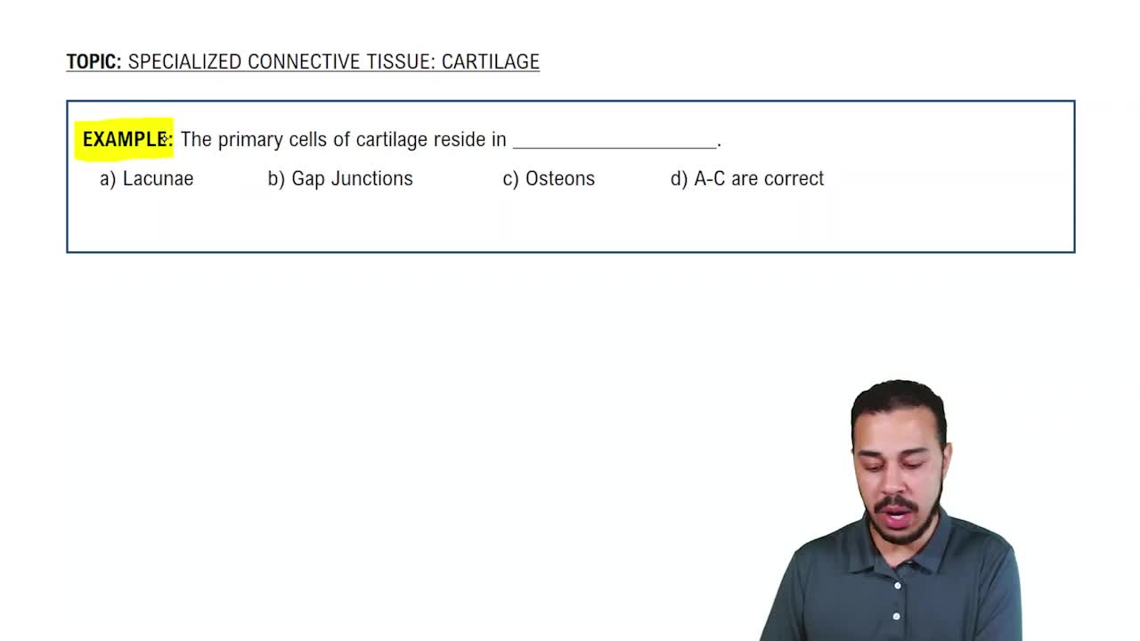
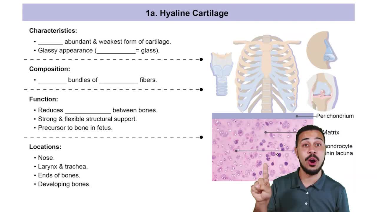
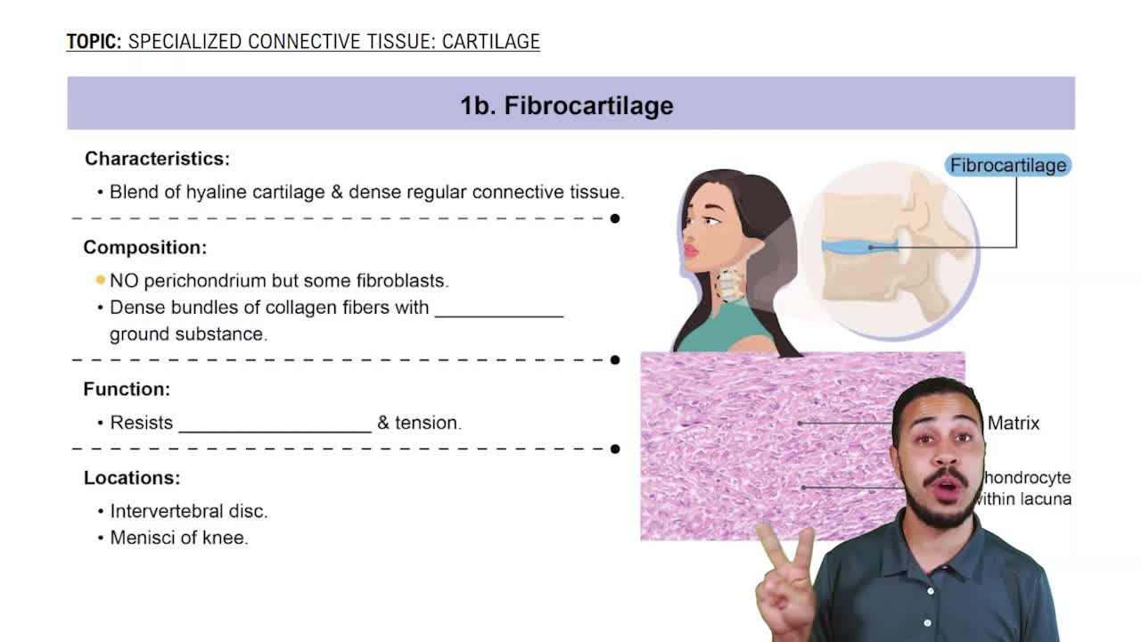
Cartilage provides flexible support, reduces friction, and absorbs shock in joints. It is avascular and composed of chondrocytes embedded in an extracellular matrix.
Hyaline: most common, found in joints and respiratory tract.
Elastic: flexible, found in ear and epiglottis.
Fibrocartilage: tough, found in intervertebral discs and pubic symphysis.
The perichondrium is a dense connective tissue layer that supports appositional growth by adding new cartilage cells to the surface.
Interstitial growth occurs from within the cartilage by division of chondrocytes, expanding the cartilage matrix internally.
Bones provide support, protection, movement, mineral storage, blood cell production, and energy storage.
Bones are classified as axial or appendicular by location; long, short, flat, irregular by shape; and composed of compact and spongy bone tissue.
Flat bones have a sandwich structure: two layers of compact bone enclosing a middle layer of spongy bone called the diploë.
Compact bone is dense and forms the outer layer of bones, providing strength and protection.
Spongy bone is porous and contains trabeculae, which are lattice-like structures that support bone marrow and reduce bone weight.
Diaphysis is the shaft of a long bone; epiphysis is the expanded end that forms joints.
The epiphyseal plate is a cartilage growth plate in children; it ossifies into the epiphyseal line after growth ends.
The medullary cavity is the central hollow space in the diaphysis containing bone marrow.
Articular cartilage covers bone ends at joints, reducing friction and absorbing shock.
The periosteum is a connective tissue membrane covering bone surfaces, containing nerves and blood vessels, and aiding in growth and repair.
The endosteum lines the medullary cavity and contains bone-forming cells.
Nutrient foramen are openings for blood vessels; perforating fibers anchor the periosteum to the bone.
Red marrow produces blood cells and is found in infants' medullary cavities and adult flat bones.
Yellow marrow stores fat and is found in adult long bone medullary cavities.
Bone cells include osteoblasts (build bone), osteocytes (maintain bone), osteoclasts (resorb bone), all derived from mesenchymal stem cells or hematopoietic lineage.
Compact bone is organized into osteons; spongy bone has trabeculae without osteons, supporting marrow and resisting stress.
Bone matrix is organic (collagen for flexibility) and inorganic (hydroxyapatite for hardness).
Endochondral ossification is bone formation from cartilage, essential for long bone growth.
The epiphyseal plate allows bones to lengthen during childhood by cartilage proliferation and ossification.
The periosteum contributes to bone thickness by adding new bone layers on the surface.
Bone remodeling maintains bone strength and mineral balance, controlled by hormones and mechanical stress.
Fracture repair involves hematoma formation, fibrocartilaginous callus, bony callus formation, and bone remodeling.
Osteoporosis is bone loss due to increased resorption or decreased formation, leading to fragile bones.
Paget’s disease causes excessive and disorganized bone remodeling, resulting in weak, deformed bones.