 뒤로
Anatomy & Physiology: Bones and Skeletal Tissues
 3:56

이 집합의 용어 (28)
Axial skeleton forms the long axis of the body including the skull, vertebral column, and rib cage. Appendicular skeleton includes bones of the upper and lower limbs and girdles attaching limbs to the axial skeleton.
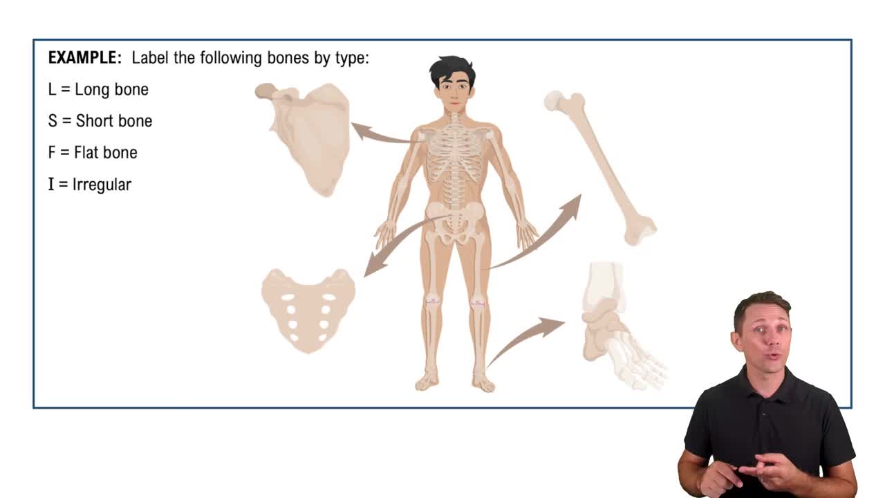
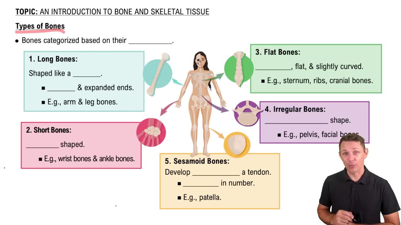
1. Long bones: longer than wide, mostly limb bones.
2. Short bones: cube-shaped, e.g., wrist and ankle bones.
3. Flat bones: thin, flat, slightly curved, e.g., sternum, ribs.
4. Irregular bones: complicated shapes, e.g., vertebrae, hip bones.
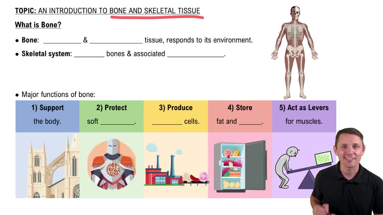
1. Support for body and soft organs.
2. Protection of brain, spinal cord, and organs.
3. Movement as levers for muscles.
4. Mineral and growth factor storage.
5. Blood cell formation (hematopoiesis).
6. Triglyceride (fat) storage.
7. Hormone production (osteocalcin).
Bones contain osseous (bone) tissue, nervous tissue, cartilage, fibrous connective tissue, muscle cells, and epithelial cells in blood vessels.
Compact bone is dense, smooth, and solid forming the outer layer. Spongy bone consists of a honeycomb of trabeculae with spaces filled with red or yellow marrow.
They consist of thin plates of spongy bone (diploë) covered by compact bone, sandwiched between connective tissue membranes (periosteum outside, endosteum inside), with scattered bone marrow and hyaline cartilage at joints.
A long bone has a diaphysis (shaft), epiphyses (ends), and membranes. The diaphysis is compact bone surrounding a medullary cavity with yellow marrow. Epiphyses have compact bone outside and spongy bone inside, covered by articular cartilage.
Periosteum: double-layered membrane covering external bone surfaces except joints, with fibrous and osteogenic layers.
Endosteum: delicate membrane covering internal bone surfaces and trabeculae, containing osteogenic cells.
Red marrow is found in trabecular cavities of spongy bone and diploë of flat bones. It is the site of hematopoiesis (blood cell formation).
1. Osteogenic cells: stem cells that differentiate.
2. Osteoblasts: bone-forming cells.
3. Osteocytes: mature bone cells maintaining matrix.
4. Bone-lining cells: maintain matrix.
5. Osteoclasts: bone-resorbing cells.
Mature bone cells in lacunae that maintain bone matrix and act as stress sensors, communicating with osteoblasts and osteoclasts for remodeling.
An osteon is the structural unit of compact bone, a cylinder with concentric lamellae containing collagen fibers in alternating directions, providing strength and resistance to twisting.
Central (Haversian) canal contains blood vessels and nerves.
Perforating (Volkmann’s) canals connect central canals.
Canaliculi are tiny canals connecting lacunae for nutrient and waste exchange.
Interstitial lamellae fill gaps between osteons or are remnants of old osteons.
Circumferential lamellae encircle the entire diaphysis beneath the periosteum, helping resist twisting.
Spongy bone is organized along lines of stress with trabeculae containing lamellae and osteocytes interconnected by canaliculi, providing strength and housing capillaries in the endosteum.
Organic: osteogenic cells, osteoblasts, osteocytes, bone-lining cells, osteoclasts, and osteoid (collagen and ground substance).
Inorganic: hydroxyapatites (calcium phosphate crystals) providing hardness and resistance to compression.
Ossification is bone tissue formation. The bony skeleton begins forming in the 2nd month of development, with postnatal growth continuing until early adulthood.
Endochondral: bone replaces hyaline cartilage, forming most bones below skull.
Intramembranous: bone develops from fibrous membranes, forming flat bones like skull and clavicles.
1. Bone collar forms around diaphysis.
2. Cartilage calcifies and cavities form.
3. Periosteal bud invades.
4. Diaphysis elongates, medullary cavity forms.
5. Epiphyses ossify.
Length grows by interstitial growth at the epiphyseal plate.
Width grows by appositional growth with osteoblasts adding bone externally and osteoclasts resorbing internally.
Growth hormone stimulates epiphyseal plate activity.
Thyroid hormone modulates growth hormone.
Testosterone and estrogens promote adolescent growth spurts and epiphyseal plate closure.
Bone remodeling is the continuous recycling of bone involving osteoblasts (bone deposit) and osteoclasts (bone resorption) at periosteal and endosteal surfaces.
Osteoclasts secrete lysosomal enzymes and acids that digest bone matrix, converting calcium salts to soluble forms, and phagocytize debris before undergoing apoptosis.
Triggers may include mechanical signals, increased calcium and phosphate ion concentrations, matrix proteins, and alkaline phosphatase enzyme for mineralization.
Parathyroid hormone (PTH) increases blood calcium by stimulating osteoclasts.
Calcitonin lowers blood calcium at high doses but has minor effects normally.
Wolff’s law states bones grow or remodel in response to mechanical stress, becoming thicker where stresses are greatest and forming trabeculae along lines of stress.
1. Hematoma formation.
2. Fibrocartilaginous callus formation.
3. Bony callus formation.
4. Bone remodeling.
Osteoporosis results from bone resorption exceeding deposit, often in postmenopausal women due to low estrogen. Prevention includes calcium and vitamin D intake, weight-bearing exercise, and avoiding smoking and excessive alcohol.