 뒤로
Anatomy & Physiology: Bones, Skeleton, and Joints
 3:56

이 집합의 용어 (73)
Dense bone tissue forming the outer layer of bones, providing strength and structure.
Found between the epiphysis and diaphysis of a long bone, marking the site of former growth plate.
Located in the medullary cavity of long bones; primarily composed of fat.
Found on the ends of bones at joints, providing a smooth, lubricated surface for articulation.
The lining of the marrow cavity inside bones.
Cells that dissolve bone matrix during bone resorption.
Concentric layers of bone matrix in compact bone.
Small channels radiating through bone matrix that connect osteocytes.
Cells responsible for building new bone matrix.
Cartilage cells responsible for early stages of endochondral ossification.
Bone growth pattern where new matrix is laid down on the surface, increasing width.
Structure indicating the end of longitudinal bone growth.
Area of longitudinal bone growth in children and adolescents.
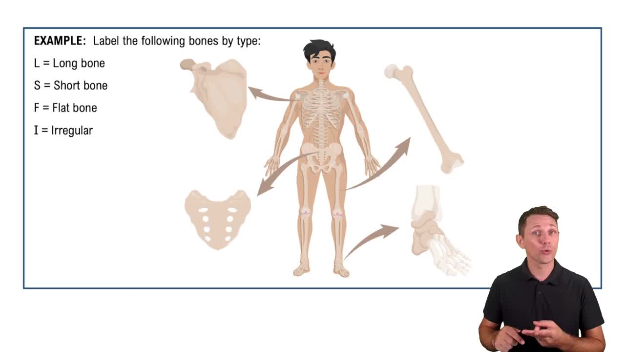
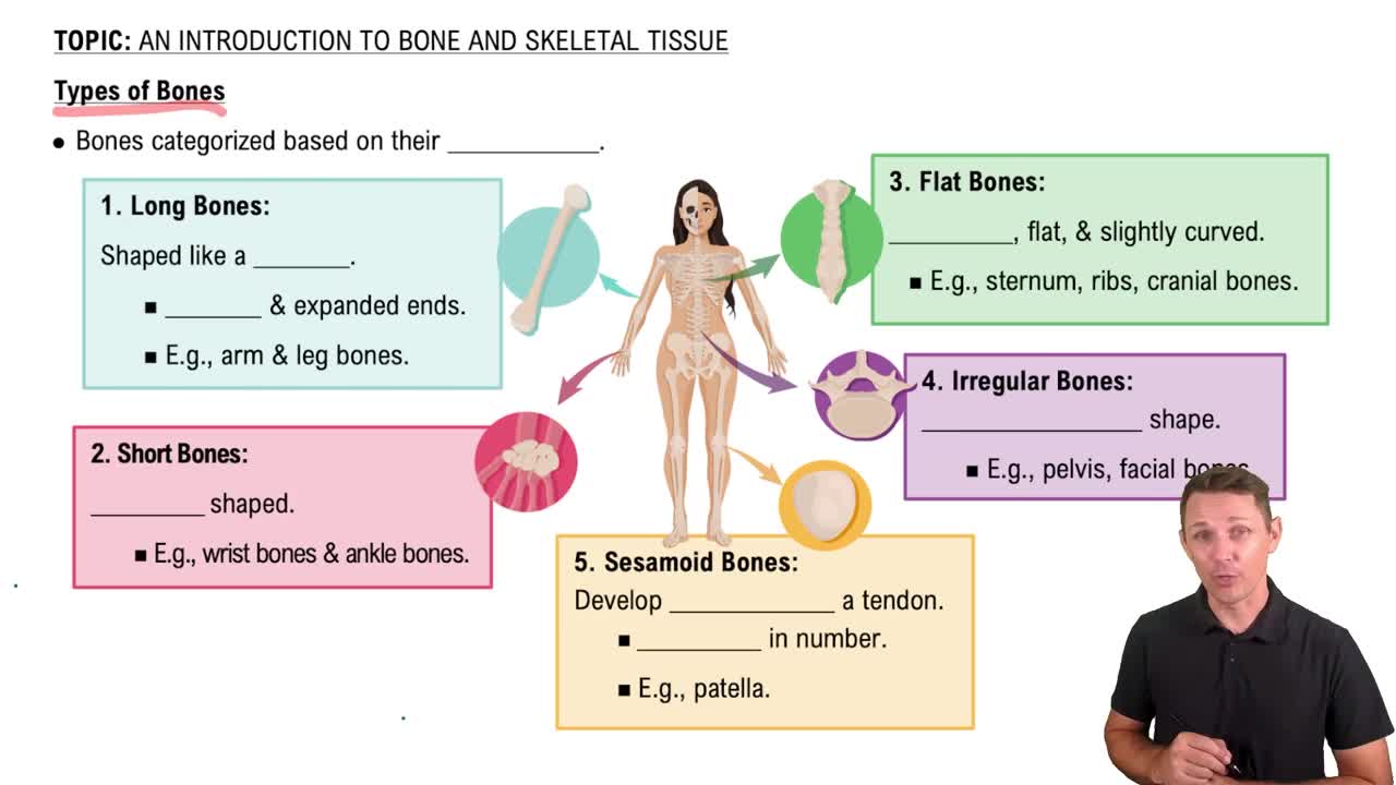
Radius is an example of a long bone.
Carpals are examples of short bones.
Patella is an example of a sesamoid bone.
Scapula is an example of a flat bone.
Hip bones are examples of irregular bones.
The formation of blood cells within the red marrow cavities of certain bones.
The organic part of the bone matrix, composed mainly of collagen fibers.
Mineral compound making up about 65% of bone mass, providing hardness.
The structural unit of compact bone, containing osteocytes, lamellae, and a central canal.
Contains a large percentage of fat.
Osteoblasts secrete the bone matrix.
Hyaline cartilage is the forerunner tissue of long bones in the embryo.
Sites where ossification of the ends of long bones occurs.
Hyaline cartilage is the most abundant type.
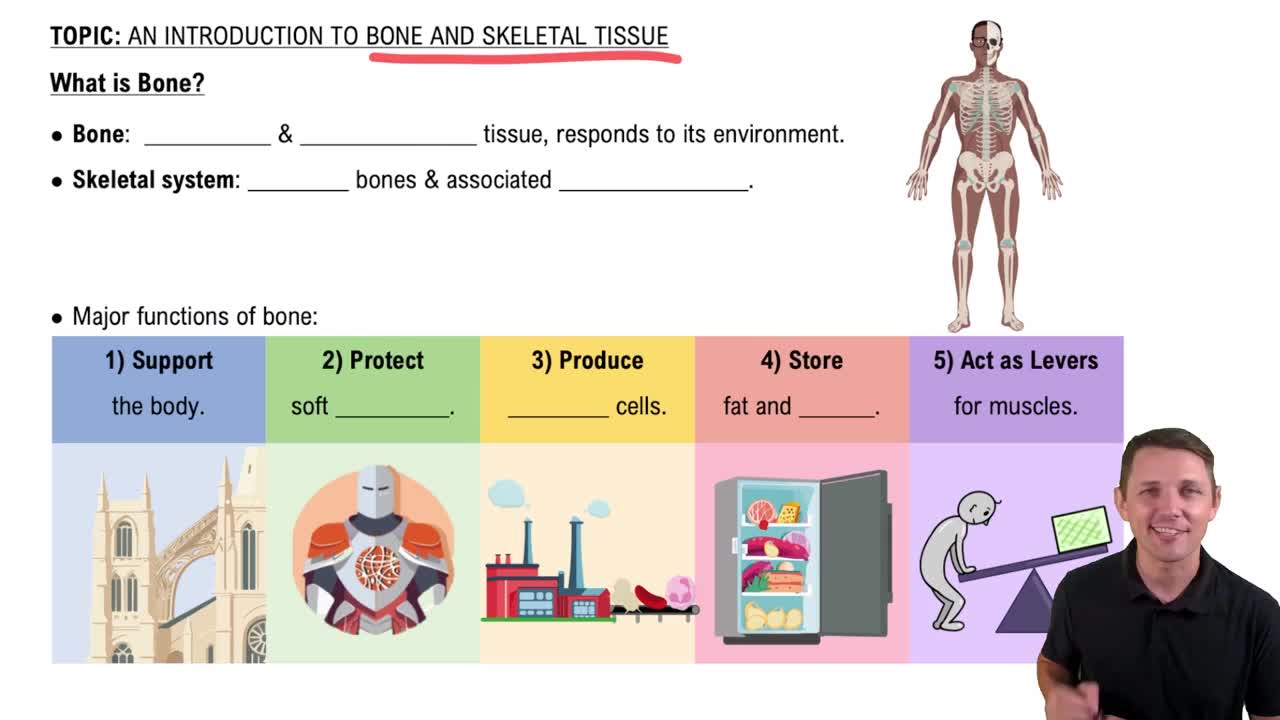
Include support, mineral storage, and blood cell production; not communication.
Contains blood vessels and nerve fibers within each osteon.
Small spaces where osteocytes reside.
Bone growth in width by adding new matrix to the surface.
Osteoclasts perform bone resorption.
Develop within fibrous membranes (intramembranous ossification).
Secretion of new matrix on the external face of existing cartilage.
Chondrocytes divide and secrete matrix, growing cartilage from within.
Made up of trabeculae, a lattice-like network.
The process of bone formation.
Occurs by interstitial growth of the epiphyseal plates during youth.
Immovable joints uniting all skull bones except the mandible.
Ribs 1-7 that attach directly to the sternum via costal cartilage.
Articulates with the transverse process of a vertebra.
Forms part of the nasal septum.
There are seven cervical vertebrae.
Does not have a body; supports the skull.
The ilium forms the largest part of the coxal bone.
Connects the two parietal bones of the skull.
Enhance voice resonance and lighten the skull.
Scoliosis is an abnormal lateral curvature, often in thoracic region.
The lumbar region bears the most body weight.
Includes ilium, ischium, and pubis; excludes the sacrum.
The fibula is not weight bearing.
The calcaneus is the heel bone.
Only cervical vertebrae have transverse foramina.
A multiaxial joint allowing movement in multiple planes.
Allows uniaxial rotation around a single axis.
Elbow joint between humerus and ulna acts as a hinge.
Allows gliding movements between flat bone surfaces.
Has concave and convex surfaces; example is thumb carpometacarpal joint.
Allows biaxial movement; example is wrist joint.
Based on the amount of movement permitted at the joint.
Movement turning the palm from posterior to anterior.
Flexion of the ankle bringing the top of the foot toward the shin.
Based on the binding material and presence or absence of a joint cavity.
Include sutures, syndesmoses, and gomphoses.
A fibrocartilaginous joint permitting slight movement.
A cartilaginous joint where hyaline cartilage unites bones.
Intercarpal and intertarsal joints.
Movement of a limb away from the body's midline along the frontal plane.
Movements that occur only at the feet.
A multiaxial synovial joint allowing movement in multiple planes.
Include synovial cavity, articular cartilage, synovial membrane, and ligaments.
Change the angle between two bones by increasing or decreasing it.
The trapezium of the carpal bone and the thumb's metacarpal form a saddle joint.