 뒤로
Anatomy & Physiology: Cardiac System Key Concepts
 3:55

이 집합의 용어 (37)
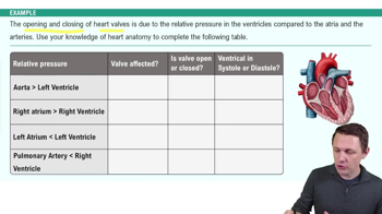
Valves between atria and ventricles that prevent backflow of blood during ventricular contraction.
An irregular heartbeat caused by abnormal electrical activity in the heart.
A bundle of specialized cardiac muscle fibers that conduct impulses from the AV node to the bundle branches.
A cluster of cells that delays the electrical impulse from the atria before passing it to the ventricles.
The upper chambers of the heart that receive blood returning to the heart.
A heart rate that is slower than normal, typically below 60 beats per minute.
Pathways that conduct electrical impulses from the bundle of His to the Purkinje fibers in the ventricles.
The sequence of contraction (systole) and relaxation (diastole) of the heart chambers during one heartbeat.
Involuntary contractions of the heart muscle that pump blood through the heart and circulatory system.
The volume of blood the heart pumps per minute, calculated as \(CO=HR \times SV\).
Specialized heart cells that generate and conduct electrical impulses to regulate heartbeat.
A condition where the heart cannot pump blood effectively, leading to fluid buildup in tissues.
The flow of blood through the vessels that supply the heart muscle (myocardium) itself.
Blood that is low in oxygen and high in carbon dioxide, returning to the heart from the body.
The phase of the cardiac cycle when the heart muscle relaxes and chambers fill with blood.
A recording of the electrical activity of the heart used to detect heart abnormalities.
The thin, smooth inner lining of the heart chambers and valves.
The outer layer of the heart wall that also forms part of the pericardium.
Rapid, irregular, and uncoordinated contractions of the heart muscle, impairing effective pumping.
The tough, outermost layer of the pericardium that protects and anchors the heart.
The number of heartbeats per minute.
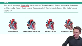
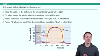
Sounds produced by the closing of heart valves during the cardiac cycle, commonly described as "lub-dub."
A network of specialized cardiac cells that initiate and distribute electrical impulses to coordinate heartbeats.
Also known as a heart attack; occurs when blood flow to part of the heart muscle is blocked, causing tissue damage.
The thick, muscular middle layer of the heart wall responsible for contraction.
Blood rich in oxygen, pumped from the lungs to the body by the heart.
The ECG wave representing atrial depolarization, which triggers atrial contraction.
The path of blood flow from the heart to the lungs and back, for oxygenation.
The ECG wave representing ventricular depolarization, leading to ventricular contraction.
The heart's natural pacemaker located in the right atrium that initiates the heartbeat.
The amount of blood ejected by a ventricle with each heartbeat.
Specialized fibers that rapidly conduct impulses throughout the ventricles to coordinate contraction.
The path of blood flow from the heart to the body tissues and back.
The phase of the cardiac cycle when the heart muscle contracts to pump blood out.
The ECG wave representing ventricular repolarization, which precedes ventricular relaxation.
A heart rate that is faster than normal, typically above 100 beats per minute.
The lower chambers of the heart that pump blood out to the lungs and body.