 뒤로
Anatomy & Physiology: Cardiovascular System and Blood Vessels
 6:13

이 집합의 용어 (49)
The cardiovascular system transports blood, delivering oxygen and nutrients to tissues and removing waste products.
The heart acts as a pump to circulate blood through the pulmonary and systemic circuits.
The heart is located in the mediastinum, between the lungs, posterior to the sternum, and above the diaphragm.
The heart pumps blood through two circuits: the right side pumps deoxygenated blood to the lungs (pulmonary circuit), and the left side pumps oxygenated blood to the body (systemic circuit).
Pericardium layers: fibrous pericardium and serous pericardium (parietal and visceral layers). Heart wall layers: epicardium, myocardium, and endocardium.
The pericardium protects the heart and anchors it; serous fluid reduces friction between pericardial layers during heartbeats.
Right atrium, right ventricle, left atrium, and left ventricle.
Right chambers contain deoxygenated blood; left chambers contain oxygenated blood.
Include superior and inferior vena cava, pulmonary trunk, pulmonary veins, and aorta.
Atrioventricular valves (tricuspid and bicuspid/mitral) and semilunar valves (pulmonary and aortic).
Remnant of the fetal foramen ovale, it allows blood flow between atria before birth.
Collects deoxygenated blood from the heart muscle and drains into the right atrium.
Chordae tendineae and papillary muscles prevent valve prolapse during ventricular contraction.
The pulmonary trunk carries deoxygenated blood from the right ventricle to the lungs; the pulmonary semilunar valve prevents backflow into the ventricle.
Pulmonary veins carry oxygenated blood to the left atrium; the bicuspid (mitral) valve controls blood flow to the left ventricle.
The aortic valve prevents backflow into the left ventricle; the aorta distributes oxygenated blood to the systemic circulation.
Supplies oxygenated blood to the heart muscle and removes deoxygenated blood.
Right coronary artery and left coronary artery.
Great cardiac vein, middle cardiac vein, and small cardiac vein.
Cardiac muscle is striated, branched, with intercalated discs containing gap junctions and desmosomes for synchronized contraction.
Depolarization (Na+ influx), plateau (Ca2+ influx), repolarization (K+ efflux).
Prevents tetanus, allowing the heart to relax and fill between beats.
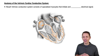
Generates and conducts electrical impulses to coordinate heart contractions.
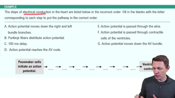
Sinoatrial (SA) node → atrioventricular (AV) node → AV bundle (Bundle of His) → bundle branches → Purkinje fibers.
It initiates the heartbeat by generating the fastest spontaneous action potentials.
Atrial depolarization preceding atrial contraction.
Ventricular depolarization preceding ventricular contraction.
Ventricular repolarization preceding ventricular relaxation.
Systole is the contraction phase; diastole is the relaxation phase of the heart chambers.
'Lup' is caused by closure of AV valves; 'dub' is caused by closure of semilunar valves.
Volume of blood pumped by each ventricle per minute.
CO = Stroke Volume (SV) × Heart Rate (HR) \(\)
Preload, contractility, and afterload affect stroke volume and cardiac output.
Increased preload stretches cardiac muscle fibers, increasing stroke volume.
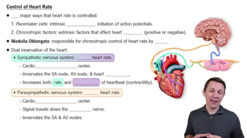
Substances that affect heart rate by altering SA node activity.
Sympathetic stimulation increases heart rate (positive chronotropic effect).
Parasympathetic stimulation decreases heart rate (negative chronotropic effect).
Arteries carry blood away from the heart; veins return blood to the heart; capillaries allow exchange of substances between blood and tissues.
Tunica intima (inner), tunica media (middle), and tunica externa (outer).
A connection between two blood vessels supplying the same region, providing alternate pathways for blood flow.
The force exerted by circulating blood on the walls of blood vessels.
The resistance to blood flow caused by friction between blood and vessel walls.
Average pressure in arteries during one cardiac cycle; MAP = diastolic pressure + 1/3 pulse pressure.
Venous return is blood flow back to the heart; assisted by skeletal muscle pump and respiratory pump.
Baroreceptors detect pressure changes; medulla oblongata controls cardiac and vasomotor centers; sympathetic and parasympathetic pathways adjust heart rate and vessel diameter.
Angiotensin II, aldosterone, ADH increase blood pressure; atrial natriuretic peptide decreases blood pressure.
Diffusion, transcytosis, and bulk flow allow substances to move between blood and tissues.
The flow of blood through body tissues to supply nutrients and remove wastes.
NFP = Net Hydrostatic Pressure (NHP) – Net Colloid Osmotic Pressure (COP).