 뒤로
Anatomy & Physiology Chapter 11: Sensory Receptors and Special Senses
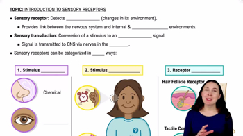
 2:47

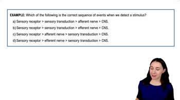
이 집합의 용어 (29)
Phasic receptors respond quickly to stimuli but adapt rapidly, signaling changes in stimulus intensity rather than constant presence.
Tonic receptors respond slowly and maintain firing as long as the stimulus is present, providing continuous information.
The specific type of stimulus to which a receptor is most sensitive and can respond, such as light for photoreceptors.
A graded electrical potential produced by sensory receptors in response to a stimulus, which can trigger an action potential if threshold is reached.
The minimum stimulus intensity required for a sensation to be consciously perceived.
The specific physical area or region where a sensory neuron can detect stimuli.
A decrease in perception of a stimulus over time due to repeated exposure, despite continued presence of the stimulus.
A neural mechanism that sharpens sensory perception by inhibiting neighboring neurons, enhancing contrast and localization.
A small central pit in the retina with a high density of cones, responsible for sharp central vision.
A photopigment found in rod cells that absorbs light and initiates the phototransduction cascade in low-light conditions.
The photopigment found in cone cells responsible for color vision under bright light.
Photoreceptors sensitive to low light levels, enabling night vision but not color detection.
Photoreceptors responsible for color vision and visual acuity in bright light.
The sensory organ within the cochlea containing hair cells that transduce sound vibrations into neural signals.
The potassium-rich fluid filling the membranous labyrinth of the inner ear, crucial for hair cell function.
The fluid surrounding the membranous labyrinth within the bony labyrinth, similar to cerebrospinal fluid.
A spiral-shaped, fluid-filled structure in the inner ear responsible for converting sound waves into nerve impulses.
Changes in the rate of rotation of the head, detected by the semicircular canals for balance.
A sensory structure in the ampulla of each semicircular canal containing hair cells that detect angular acceleration.
A gelatinous structure in the crista ampullaris that bends hair cells in response to fluid movement during head rotation.
One of the otolithic organs in the vestibule that detects horizontal linear acceleration and head position.
One of the otolithic organs in the vestibule that detects vertical linear acceleration and head position.
Structures (utricle and saccule) containing hair cells and otoliths that detect linear acceleration and gravity.
Hair-like projections on hair cells that bend in response to fluid movement, initiating sensory transduction.
Three fluid-filled canals in the inner ear that detect rotational movements of the head.
The process by which the eye's lens changes shape to focus on objects at different distances.
Rods are depolarized and continuously release neurotransmitter, signaling the absence of light.
Rods hyperpolarize, reducing neurotransmitter release, which signals the presence of light.
Convergence of multiple rods onto a single neuron increases sensitivity in low light but reduces visual acuity.