 뒤로
Anatomy & Physiology Chapter 18: Blood Study Guide
 2:36

이 집합의 용어 (26)
Transports nutrients, signaling molecules, respiratory gases, and waste products; carries hormones and immune cells; delivers oxygen and nutrients to tissues; helps regulate body temperature.
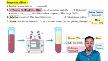
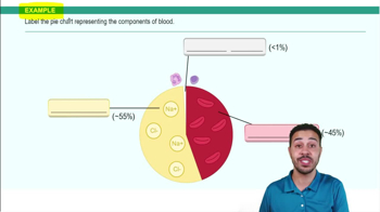
Blood is a specialized connective tissue composed of plasma (55%) and formed elements (erythrocytes, leukocytes, platelets).
Percentage of blood volume occupied by red blood cells; about 47% ± 5% in males and 42% ± 5% in females.
Thin layer between plasma and erythrocytes containing leukocytes and platelets; less than 1% of whole blood.
Albumin: maintains blood osmotic pressure; Globulins: antibodies and transport proteins; Fibrinogen: blood clotting.
Biconcave shape increases surface area; no nucleus or organelles maximizes hemoglobin space; no mitochondria prevents oxygen use by the cell.
Four amino acid chains each with an iron-binding site; iron binds oxygen and gives blood its red color when oxygenated.
Granulocytes: neutrophils, eosinophils, basophils; Agranulocytes: lymphocytes, monocytes.
Never Let Monkeys Eat Bananas = Neutrophils, Lymphocytes, Monocytes, Eosinophils, Basophils (most to least abundant).
50–70% of WBCs; 2–6 nuclear lobes; pale red/blue granules; first-line inflammatory defense and phagocytizes bacteria.
20–45% of WBCs; large dark spherical nucleus with thin cytoplasm rim; specific immune defense via T and B cells.
3–8% of WBCs; largest WBC with kidney-shaped nucleus; leaves blood to become phagocytic macrophage.
1–4% of WBCs; bilobed nucleus with large red granules; fights parasitic infections and allergic responses.
0.5–1% of WBCs; bilobed nucleus with dark purplish-black granules; releases histamine and mediates inflammation.
Fragments from megakaryocytes; plug vessel tears, release factors for clotting, vasoconstriction, and inflammation; contract to close wounds.
Found in spongy bone of axial skeleton and proximal epiphyses; actively produces new blood cells.
Located in long bones; mostly fat cells; usually dormant but can produce blood cells in emergencies.
Process of blood cell formation; about 100 billion new blood cells formed daily in red marrow.
Lymphoid stem cells produce lymphocytes; myeloid stem cells produce erythrocytes, megakaryocytes/platelets, granulocytes, and monocytes.
Proerythroblast → erythroblast → reticulocyte → erythrocyte; reticulocytes mature in circulation for 1–2 days.
Abnormally high red blood cell concentration; polycythemia vera is caused by bone marrow cancer.
Low red blood cell count or hemoglobin concentration, leading to reduced oxygen transport.
Inherited defective hemoglobin causes red blood cells to become sickle-shaped, impairing oxygen delivery.
Cancer of leukocytes classified by cell line (lymphoblastic or myeloblastic) and progression rate (acute or chronic).
Abnormally low platelet count caused by marrow damage, chemotherapy, vitamin B12 deficiency, or leukemia.
Process by which leukocytes squeeze through capillary walls to reach infection sites.