뒤로

Anatomy & Physiology: Epithelial and Connective Tissue Characteristics

컨트롤 버튼이 '내비게이션' 모드로 변경되었습니다.
1/32
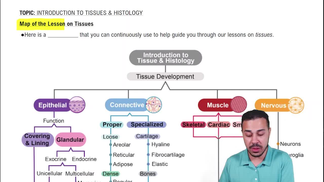
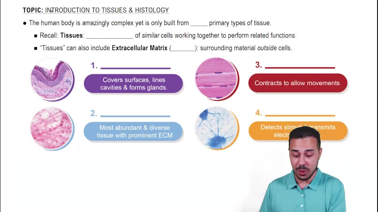
  • Five characteristics of epithelial tissue

    • Polarity: Apical and basal surfaces
    • Specialized contacts: Tight junctions and desmosomes bind cells
    • Supported by connective tissue
    • Avascular but innervated: Nourished by diffusion, supplied by nerve fibers
    • Regeneration: High capacity stimulated by loss of polarity and broken contacts
  • Simple squamous epithelial tissue structure and function

    Single layer of flattened cells with disc-shaped nuclei; allows rapid diffusion, absorption, secretion, or filtration.
  • Locations of simple squamous epithelium

    Kidney glomeruli, air sacs of lungs, lining of heart, blood vessels, and lymphatic vessels.
  • Two special types of simple squamous epithelium

    Endothelium: lining of blood vessels;

    Mesothelium: lining of body cavities.

  • Simple cuboidal epithelial tissue features

    Single layer of cube-shaped cells with large spherical nuclei; functions in secretion and absorption.
  • Locations of simple cuboidal epithelium

    Kidney tubules, ducts and secretory portions of small glands, and ovary surface.
  • Simple columnar epithelial tissue characteristics

    Single layer of tall, closely packed cells; may have microvilli or cilia; some contain mucus-secreting goblet cells.
  • Functions of simple columnar epithelium

    Absorption and secretion of mucus, enzymes, and other substances; ciliated cells propel mucus.
  • Locations of simple columnar epithelium

    Nonciliated: digestive tract, gallbladder, ducts of some glands; Ciliated: bronchi, uterine tubes, uterus.
  • Pseudostratified columnar epithelial tissue features

    Cells vary in height, appear multilayered but all contact basement membrane; nuclei at different levels.
  • Functions of pseudostratified columnar epithelium

    Secretes mainly mucus; cilia propel mucus.
  • Locations of pseudostratified columnar epithelium

    Ciliated: trachea and upper respiratory tract;

    Nonciliated: sperm-carrying ducts and ducts of large glands.

  • Stratified squamous epithelial tissue characteristics

    Two or more layers; basal cells divide and migrate upward; surface cells are squamous; provides protection.
  • Locations and types of stratified squamous epithelium

    Keratinized: epidermis of skin (dry); Nonkeratinized: moist linings of esophagus, mouth, vagina.
  • Transitional epithelial tissue features and function

    Lines hollow urinary organs; basal cells cuboidal or columnar; cells change shape when stretched to allow urine flow and storage.
  • Three characteristics of connective tissue

    • Extracellular matrix: nonliving, supports cells
    • Common origin: from mesenchyme
    • Vascularity: varies by tissue type
  • Areolar connective tissue description and function

    Gel-like matrix with collagen fibers, fibroblasts, macrophages; supports and binds tissues; cushions organs; holds interstitial fluid.
  • Locations of areolar connective tissue

    Under epithelia, packaging organs, surrounding capillaries.
  • Adipose connective tissue features and functions

    Closely packed adipocytes with large fat droplets; stores nutrients; shock absorption; insulates; supports organs.
  • Locations of adipose connective tissue

    Subcutaneous tissue, around kidneys and eyeballs, breasts, abdomen.
  • Reticular connective tissue characteristics and function

    Loose network of reticular fibers and cells; forms stroma supporting blood cells in lymphoid organs.
  • Locations of reticular connective tissue

    Lymph nodes, spleen, bone marrow.
  • Dense regular connective tissue features

    Parallel bundles of thick collagen fibers; high tensile strength; few cells; poorly vascularized.
  • Functions and locations of dense regular connective tissue

    Attaches muscles to bones or muscles, bones to bones; withstands tension in one direction; found in tendons and ligaments.
  • Dense irregular connective tissue characteristics

    Thicker collagen bundles arranged irregularly; forms sheets; resists tension from many directions.
  • Locations of dense irregular connective tissue

    Dermis of skin, fibrous joint capsules, organ coverings, submucosa of digestive tract.
  • Hyaline cartilage features and functions

    Most abundant cartilage; shiny blue glass appearance; collagen fibers form imperceptible network; supports and cushions; resists compression.
  • Locations of hyaline cartilage

    Tips of long bones, nose, trachea, larynx, embryonic skeleton, ribs cartilage.
  • Elastic cartilage characteristics and function

    Similar to hyaline but with more elastic fibers; maintains shape with flexibility.
  • Locations of elastic cartilage

    External ear and epiglottis.
  • Fibrocartilage features and function

    Matrix less firm than hyaline with thick collagen fibers; very strong; absorbs compressive shock.
  • Locations of fibrocartilage

    Intervertebral discs, pubic symphysis, knee joint discs.