 뒤로
Anatomy & Physiology Exam 3 Review
 04:06

이 집합의 용어 (26)
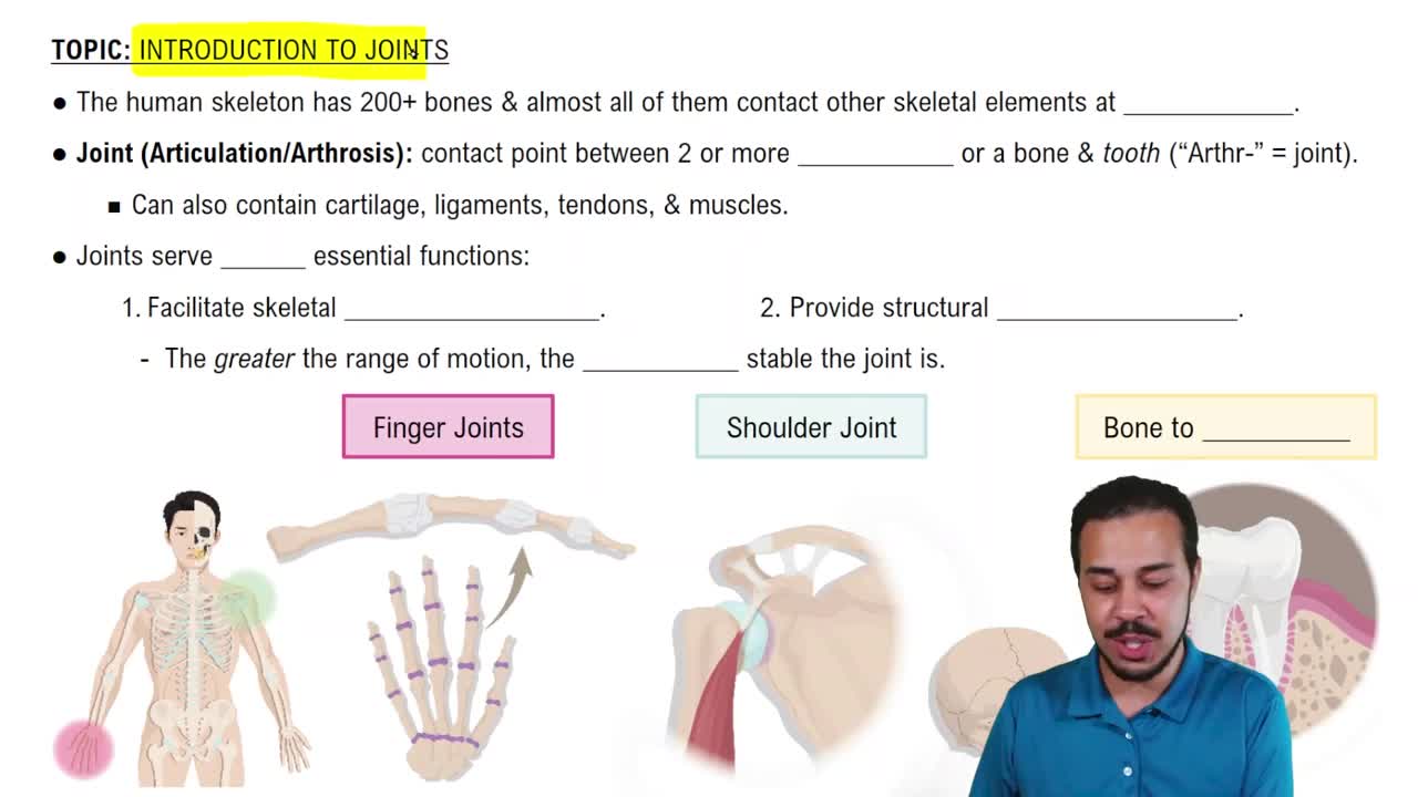
Synovial joints include the articular cartilage, synovial membrane, joint cavity, synovial fluid, ligaments, and the joint capsule.
Synovial joints allow movement in planes such as sagittal, frontal, and transverse depending on the joint type.
Flexion is the movement that decreases the angle between two bones.
Extension is the movement that increases the angle between two bones.
Abduction is the movement of a limb away from the midline of the body.
Adduction is the movement of a limb toward the midline of the body.
A sprained ankle involves stretched or torn ligaments around the ankle joint.
Tommy John surgery repairs a torn ulnar collateral ligament in the elbow.
Osteoarthritis is wear-and-tear joint degeneration; rheumatoid arthritis is an autoimmune inflammation of joints.
Articular cartilage covers bone ends in joints to reduce friction and absorb shock.
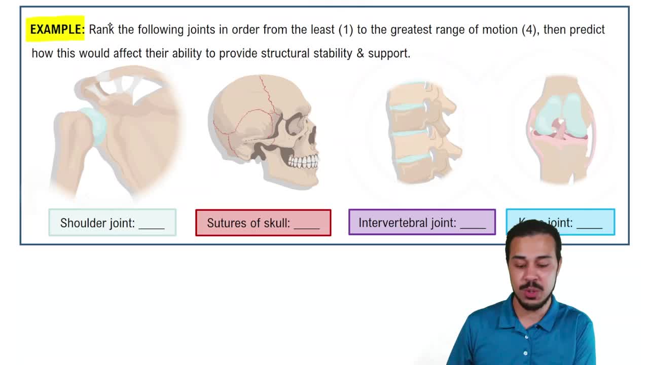
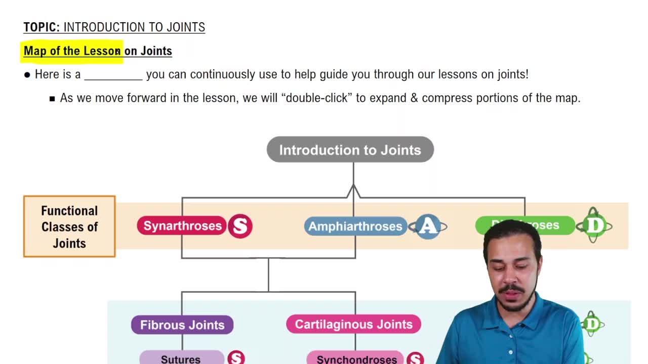
Cartilaginous joints include synchondroses (hyaline cartilage) and symphyses (fibrocartilage).
Fibrous joints include sutures, syndesmoses, and gomphoses, connected by dense connective tissue.
The patellar ligament connects the patella to the tibia and is involved in the knee-jerk reflex.
A muscle fiber contains myofibrils, sarcolemma, sarcoplasm, mitochondria, and nuclei.
A sarcomere is the functional unit of muscle contraction, defined by Z-discs and containing actin and myosin filaments.
A motor neuron transmits nerve impulses from the central nervous system to muscle fibers.
Muscle contractions include isotonic (concentric and eccentric) and isometric contractions.
Cardiac muscle is striated and involuntary; skeletal muscle is striated and voluntary; smooth muscle is non-striated and involuntary.
Muscle tissue enables movement, maintains posture, stabilizes joints, and generates heat.
Key proteins include actin, myosin, troponin, and tropomyosin.
Rigor mortis occurs due to ATP depletion, causing myosin heads to remain bound to actin.
During vigorous exercise, pyruvate is converted to lactic acid via anaerobic glycolysis.
The sliding filament model describes actin filaments sliding over myosin filaments to shorten the sarcomere during contraction.
During contraction, the sarcomere shortens, the H zone and I band narrow, while the A band remains constant.
Action potential steps: depolarization, repolarization, and hyperpolarization of the neuron membrane.
If acetylcholine (ACh) is blocked at the neuromuscular junction, muscle contraction cannot be initiated.