 뒤로
Anatomy & Physiology Exam 3 Review
 04:06

이 집합의 용어 (27)
Articular cartilage, synovial membrane, joint cavity, ligaments, and fibrous capsule are key parts of a synovial joint.
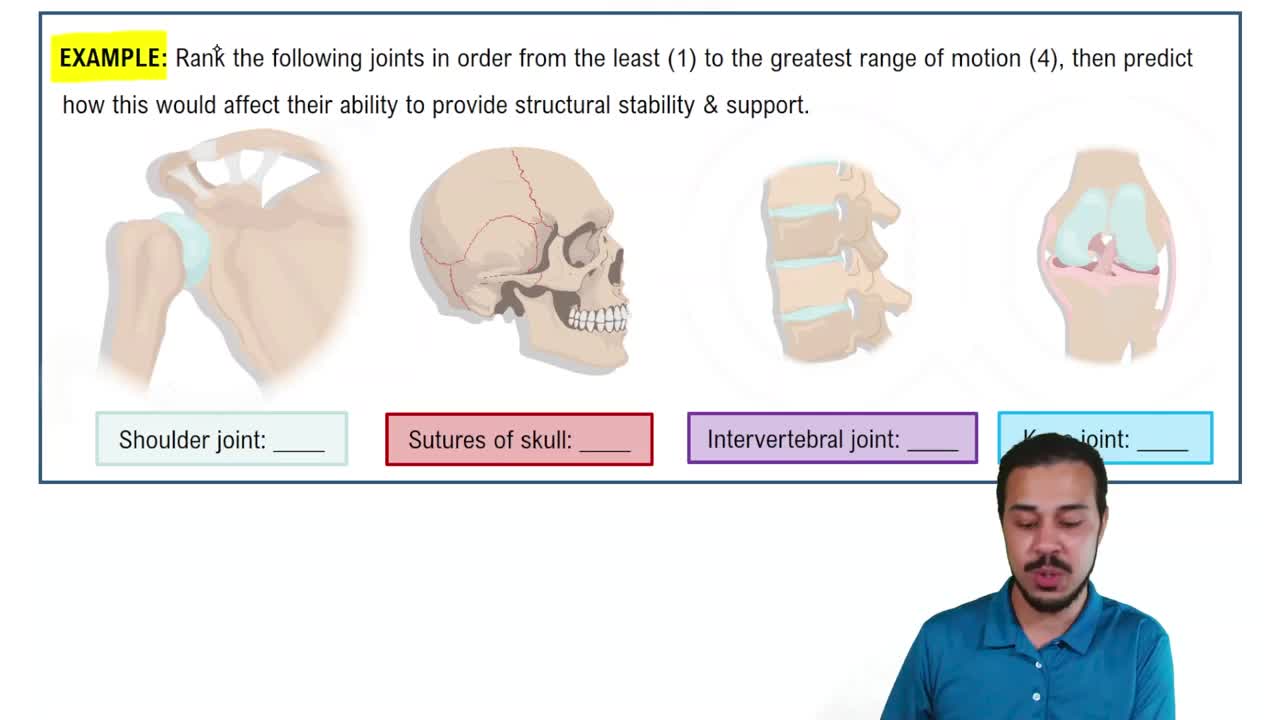
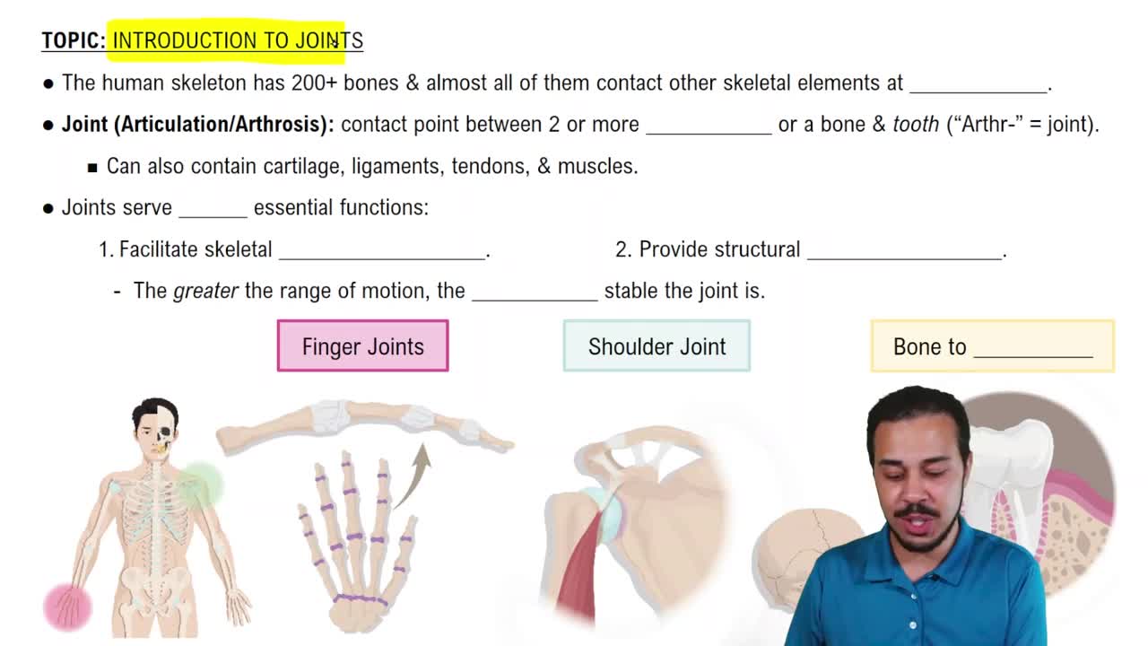
Synovial joints allow movement in uniaxial, biaxial, or multiaxial planes depending on joint type.
Flexion decreases the angle between two bones at a joint.
Extension increases the angle between two bones at a joint.
Abduction moves a limb away from the midline of the body.
Adduction moves a limb toward the midline of the body.
Damage or tearing of ligaments supporting the ankle joint.
Reconstruction of the ulnar collateral ligament in the elbow due to injury.
Inflammation of joints causing pain and stiffness.
Autoimmune disorder attacking joint linings.
Degeneration of articular cartilage leading to bone friction.
Provides a smooth, lubricated surface for joint movement and absorbs shock.
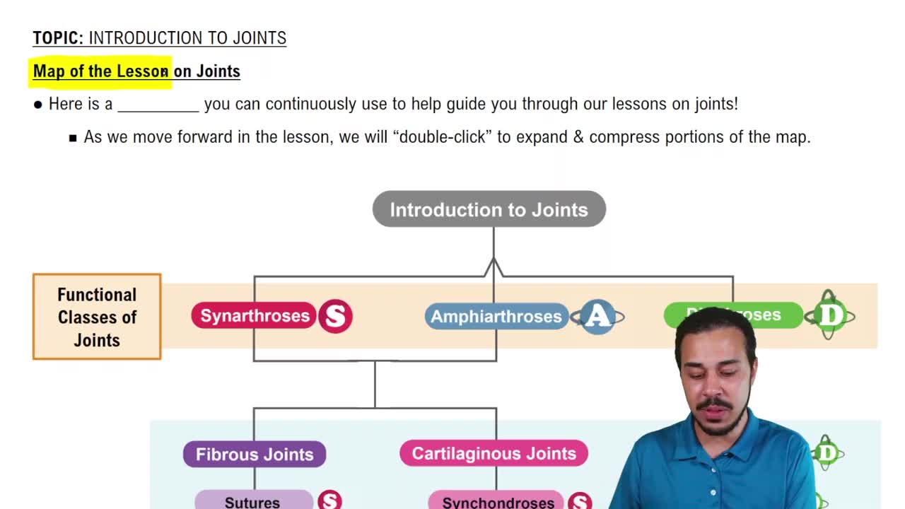
Synchondroses (hyaline cartilage) and symphyses (fibrocartilage) are main types.
Sutures, syndesmoses, and gomphoses connect bones with fibrous tissue.
Patellar ligament transmits the reflex signal causing quadriceps contraction.
Includes sarcolemma, myofibrils, sarcomeres, mitochondria, and sarcoplasmic reticulum.
A nerve cell that transmits impulses from the central nervous system to muscle fibers.
Isotonic (concentric and eccentric) and isometric contractions.
Cardiac: striated, involuntary; Skeletal: striated, voluntary; Smooth: non-striated, involuntary.
Movement, posture maintenance, heat production, and joint stabilization.
Actin, myosin, troponin, and tropomyosin.
ATP depletion prevents detachment of myosin from actin, causing muscle stiffness.
Converted to lactic acid via anaerobic glycolysis.
Actin filaments slide over myosin filaments, shortening the sarcomere during contraction.
I band and H zone shorten; A band remains constant.
Resting state, depolarization, repolarization, and hyperpolarization phases.
Muscle contraction is prevented due to lack of signal transmission.