 뒤로
Anatomy & Physiology Exam 3 Study Guide
 04:06

이 집합의 용어 (30)
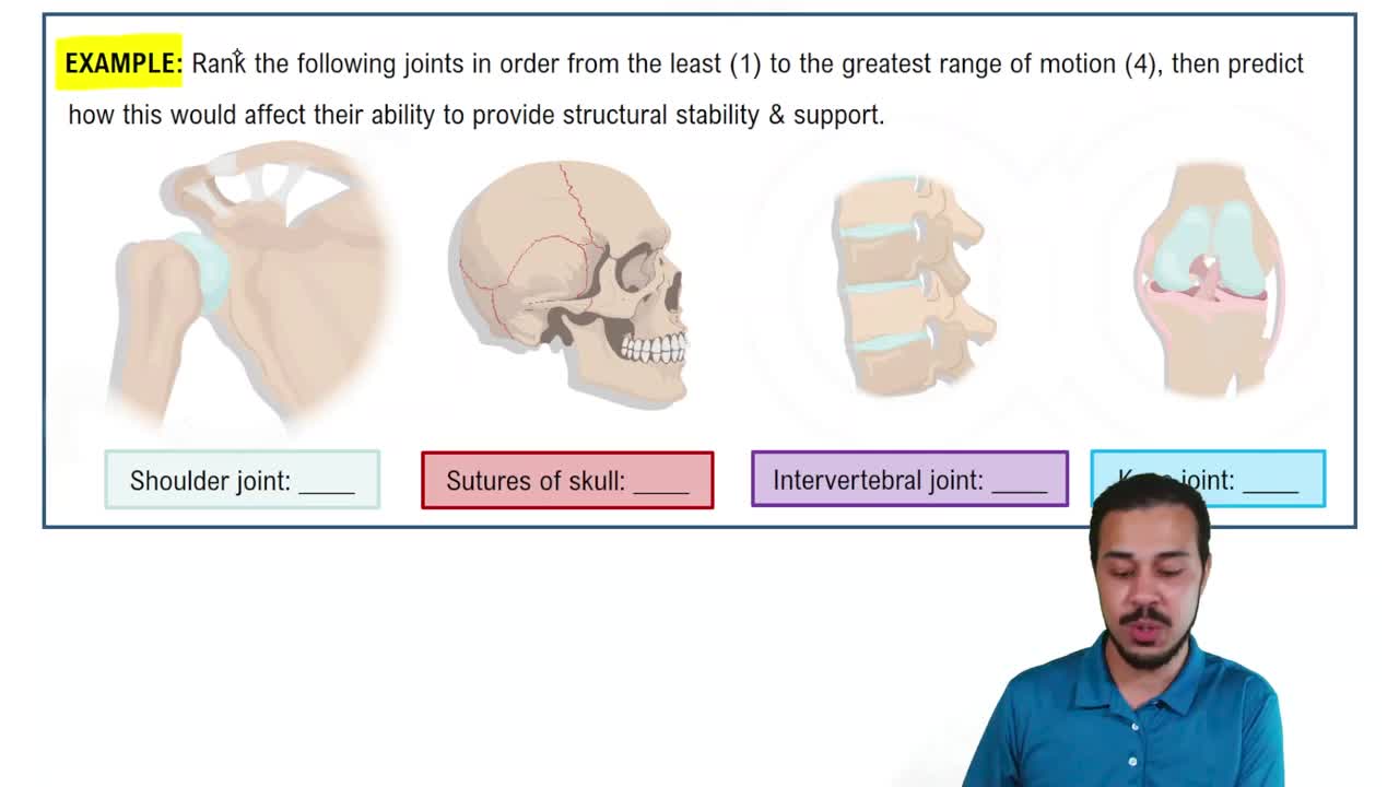
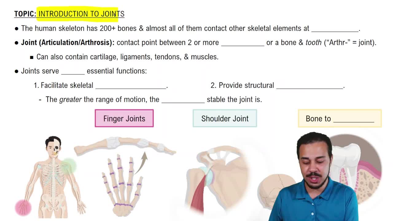
A joint is the connection point between two or more bones that allows for varying degrees of movement or stability.
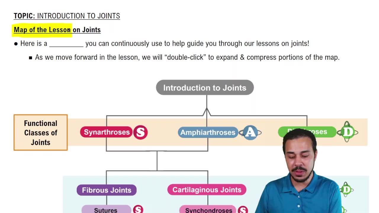
There are three main types: Fibrous, Cartilaginous, and Synovial joints, classified by their structure and movement capability.
Fibrous joints are connected by dense connective tissue, allowing little or no movement. Examples include sutures in the skull and syndesmoses.
Cartilaginous joints are connected by cartilage, allowing limited movement. Examples include intervertebral discs and the pubic symphysis.
Synovial joints have a fluid-filled joint cavity, articular cartilage, synovial membrane, and a fibrous capsule, allowing free movement.
Synovial joints differ by having a joint cavity with synovial fluid, enabling greater mobility compared to fibrous and cartilaginous joints.
The six types are: plane, hinge, pivot, condyloid, saddle, and ball-and-socket joints.
Synovial joints can be monoaxial (one plane), biaxial (two planes), or multiaxial (multiple planes).
Flexion decreases the angle between bones; extension increases the angle, returning to anatomical position.
Hyperextension is the extension of a joint beyond its normal anatomical range.
Dorsiflexion is lifting the foot upwards; plantar flexion is pointing the foot downwards.
Inversion turns the sole of the foot inward; eversion turns it outward.
Elevation raises the scapula; depression lowers it.
Upward rotation moves the scapula so the glenoid cavity faces up; downward rotation returns it to anatomical position.
Circumduction is a circular movement combining flexion, extension, abduction, and adduction.
Internal rotation turns a limb toward the body's midline; external rotation turns it away.
Levers differ by the order of fulcrum (F), load (L), and effort (E): 1st class (F between E and L), 2nd class (L between F and E), 3rd class (E between F and L).
Mechanical advantage is the ratio of output force to input force; 2nd class levers provide greatest MA, 3rd class the least.
An action potential is a rapid, temporary change in a cell's membrane potential that transmits electrical signals.
Phases include depolarization (membrane becomes positive), repolarization (returns to negative), and hyperpolarization (more negative than resting).
The sodium-potassium pump maintains ion gradients by pumping 3 Na+ out and 2 K+ in, essential for resting potential and action potentials.
The absolute refractory period prevents new APs; the relative refractory period allows APs with stronger stimulus.
A myofibril is a contractile organelle in muscle cells; myofilaments are protein filaments (actin and myosin) within myofibrils.
The sarcomere is the functional unit of muscle contraction, defined by Z discs, with A bands, I bands, and M lines organizing filaments.
The sliding filament theory explains muscle contraction by actin filaments sliding over myosin, shortening the sarcomere.
Troponin binds calcium to move tropomyosin off actin's binding sites, allowing myosin to bind and contract muscle.
Includes the motor neuron terminal, synaptic cleft, and muscle fiber membrane; acetylcholine is released to trigger muscle contraction.
Three types: isotonic concentric (muscle shortens), isotonic eccentric (muscle lengthens), and isometric (muscle length stays same).
Skeletal is voluntary and striated; cardiac is involuntary, striated with intercalated discs; smooth is involuntary, non-striated with gap junctions.
Calcium binds calmodulin, activating myosin light chain kinase, which phosphorylates myosin to enable smooth muscle contraction.