 뒤로
Anatomy & Physiology Exam 3 Study Guide
 04:06

이 집합의 용어 (31)
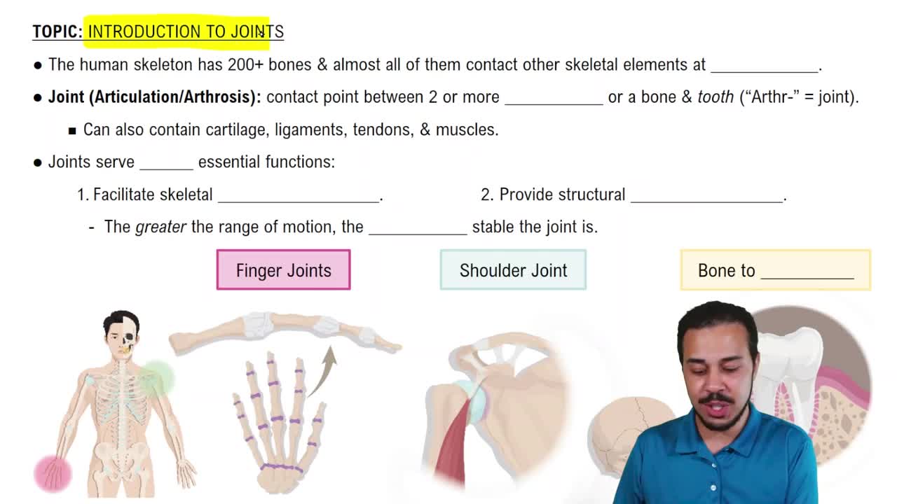
Articulations are joints where two or more bones meet, allowing for movement and providing mechanical support.
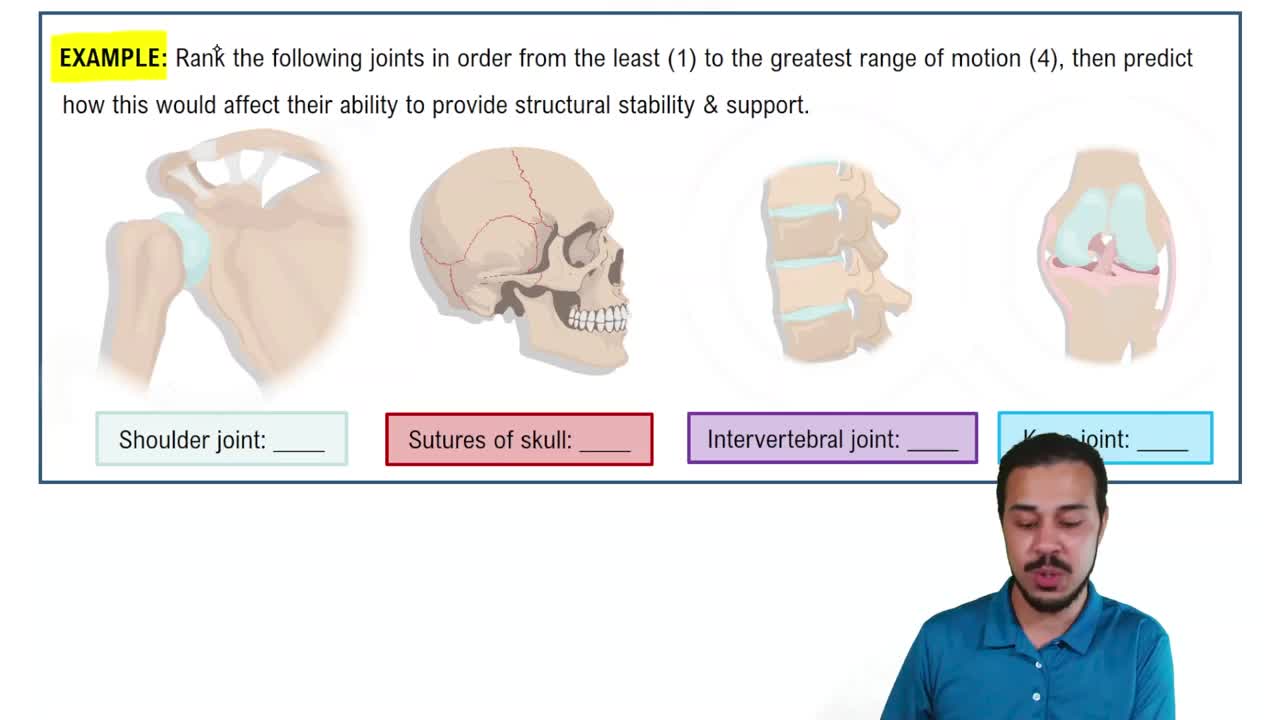
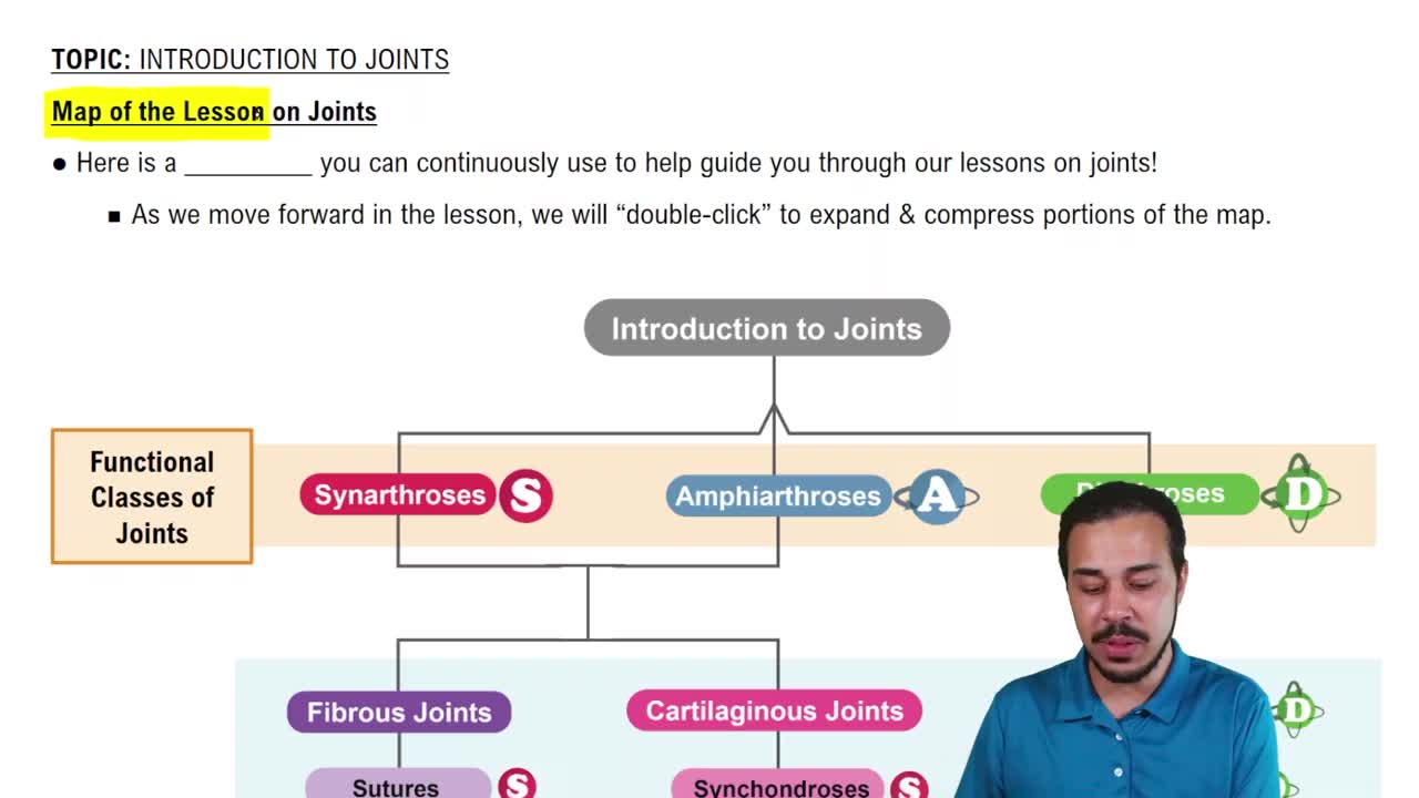
Joints are classified structurally as fibrous, cartilaginous, or synovial based on the type of connective tissue binding the bones.
Fibrous joints include sutures in the skull (immovable) and syndesmoses like the distal tibiofibular joint (slightly movable).
Functional classification groups joints by movement: synarthroses (immovable), amphiarthroses (slightly movable), and diarthroses (freely movable).
Cartilaginous joints connect bones with cartilage, allowing limited movement and lacking a joint cavity.
Synchondroses (e.g., epiphyseal plates) and symphyses (e.g., pubic symphysis) are the two main types.
Synovial joints have a joint cavity filled with synovial fluid, articular cartilage, a fibrous capsule, synovial membrane, ligaments, and often bursae or tendon sheaths.
Features include articular cartilage, joint cavity, articular capsule, synovial fluid, ligaments, and nerves and blood vessels.
Bursae are fluid-filled sacs reducing friction between tissues; tendon sheaths are elongated bursae surrounding tendons to reduce friction during movement.
Joint stability is enhanced by muscle tone, ligaments, and the shape of articulating surfaces.
Movements include flexion, extension, abduction, adduction, rotation, and circumduction.
Types are plane, hinge, pivot, condylar, saddle, and ball-and-socket joints.
The humerus and scapula form the shoulder joint, a ball-and-socket joint allowing wide range of motion.
The elbow is a hinge joint between the humerus, radius, and ulna, allowing flexion and extension.
The hip joint is a ball-and-socket joint between the femur and the acetabulum of the pelvis.
The temporomandibular joint (TMJ) connects the jawbone to the skull, allowing hinge and gliding movements.
The 3 C’s are cruciate ligament injury, cartilage (meniscus) tear, and collateral ligament injury.
Osteoarthritis is degenerative; rheumatoid arthritis is autoimmune; gouty arthritis results from uric acid crystals.
Lyme disease is caused by a tick-borne bacterium, leading to joint inflammation and arthritis if untreated.
Lower incidence is due to fewer tick vectors and different ecological conditions on the West Coast.
Muscle types are skeletal (voluntary), cardiac (heart), and smooth (involuntary, walls of organs).
Muscle tissue is excitability, contractility, extensibility, and elasticity.
Functions include movement, posture maintenance, joint stabilization, and heat generation.
A skeletal muscle is composed of muscle fibers bundled by connective tissue sheaths: endomysium, perimysium, and epimysium.
Myofibrils contain contractile filaments; sarcoplasmic reticulum stores calcium; T tubules transmit action potentials.
During contraction, actin filaments slide over myosin filaments, shortening the sarcomere without changing filament length.
Steps: cross bridge formation, power stroke, cross bridge detachment, and cocking of myosin head.
Action potential triggers acetylcholine release, which binds receptors, causing depolarization and muscle fiber excitation.
Myasthenia gravis is an autoimmune disorder where antibodies block acetylcholine receptors, causing muscle weakness.
Rigor mortis occurs due to ATP depletion after death, preventing cross bridge detachment and causing muscle stiffness.
ATP is regenerated by creatine phosphate, anaerobic glycolysis, and aerobic respiration.