 뒤로
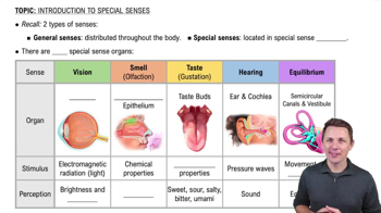
Anatomy & Physiology: Eye and Ear Structures and Functions
 5:05

이 집합의 용어 (33)
is the transparent front layer of the eye that refracts light to help focus it onto the retina.
is the white, tough outer layer of the eye that provides protection and maintains eye shape.
controls the diameter of the pupil, regulating the amount of light entering the eye to optimize vision.
transmits visual information from the retina to the brain for image processing.
produces tears that lubricate and protect the surface of the eye.
elevates the eye and helps rotate it medially.
depresses the eye and assists in medial rotation.
moves the eye medially (toward the nose) to focus on near objects. ADDucts
moves the eye laterally (away from the nose) to widen the field of view. ABduct
rotates the eye downward and laterally, aiding in complex eye movements.
rotates the eye upward and laterally, helping with eye alignment and tracking.
is the clear gel filling the eye's interior, maintaining its shape and supporting the retina.
focuses light onto the retina by changing shape to adjust for near or far vision (accommodation).
is the opening in the iris that controls the amount of light entering the eye.
enlarge the pupil to allow more light in low-light conditions, improving night vision.
reduce pupil size to limit light entry and protect the retina in bright conditions.
controls lens shape for focusing and produces aqueous humor to nourish the eye.
contains photoreceptor cells that convert light into neural signals for vision.
is the blind spot where the optic nerve exits the eye; it lacks photoreceptors.
supplies blood to the retina; the Tapetum Lucidum enhances night vision by reflecting light within the eye.
is the external ear structure that collects and directs sound waves into the ear canal.
is the ear canal that channels sound waves to the tympanic membrane.
(eardrum) vibrates in response to sound waves, transmitting them to the middle ear bones.
equalizes air pressure between the middle ear and the throat to maintain eardrum function.
converts sound vibrations into electrical signals for hearing via hair cells.
is the first middle ear bone that transmits vibrations from the tympanic membrane to the incus.
is the middle ear bone that passes vibrations from the malleus to the stapes.
transmits vibrations from the incus to the oval window of the inner ear.
is the membrane that receives vibrations from the stapes and transmits them into the cochlea fluid.
allows fluid movement within the cochlea by bulging outward as vibrations enter.
detects linear acceleration and head position to help maintain balance.
detect rotational movements of the head to aid in balance and spatial orientation.
(vestibulocochlear nerve) transmits auditory and balance information from the ear to the brain.