Skip to main content
Anatomy & Physiology
나의 코스
배우기
시험 준비
AI 튜터
학습 가이드
교과서적 해결책
플래시카드
탐험해 보세요
앱을 사용해 보세요
나의 코스
배우기
시험 준비
AI 튜터
학습 가이드
교과서적 해결책
플래시카드
탐험해 보세요
앱을 사용해 보세요
뒤로
Anatomy & Physiology II: Blood - Key Concepts
카드를 뒤집기 위해 탭할 수 있습니다.
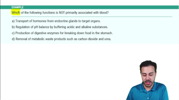
Primary functions of blood
카드를 뒤집기 위해 탭할 수 있습니다.
👆
Primary functions of blood
Transport
of O2, nutrients, wastes, and hormones;
Regulation
of body temperature, pH, and fluid volume;
Protection
against blood loss and infection.
진행 상황 추적
컨트롤 버튼이 '내비게이션' 모드로 변경되었습니다.
1/29
추천 영상
VideoThumbView.guidedCourse
2:36
Introduction to Blood Example 1
11363
views
334
rank
VideoThumbView.guidedCourse
7:49
Composition of Blood
11908
views
337
rank
VideoThumbView.guidedCourse
1:55
Introduction to Blood Example 2
9380
views
225
rank
이 집합의 용어 (29)
하이드의 정의
Primary functions of blood
Transport
of O2, nutrients, wastes, and hormones;
Regulation
of body temperature, pH, and fluid volume;
Protection
against blood loss and infection.
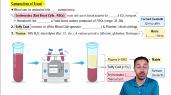
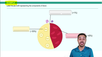
Composition of blood
Blood is a connective tissue with a fluid matrix called
plasma
and living cells called
formed elements
(erythrocytes, leukocytes, platelets).
Hematocrit
The percentage of blood volume composed of
erythrocytes
, normally about 45%.
Physical characteristics of blood
Sticky, opaque fluid with metallic taste; color varies with oxygen content; pH 7.35–7.45; volume ~5 L in adults.
Major plasma proteins and their functions
Albumin
(carrier, buffer, osmotic pressure),
globulins
(immune function), and
fibrinogen
(clotting).
Erythrocyte structure
Biconcave, anucleate cells filled with hemoglobin; flexible due to spectrin; optimized for gas transport.
Hemoglobin composition
Four polypeptide chains (2 alpha, 2 beta) each with an iron-containing heme group that binds oxygen.
Erythropoiesis
Process of RBC formation in red bone marrow, taking about 15 days from stem cell to mature erythrocyte.
Regulation of erythropoiesis
Controlled by erythropoietin (EPO) released by kidneys in response to hypoxia; testosterone enhances EPO production.
Dietary requirements for RBC production
Adequate amino acids, iron (stored as ferritin), vitamin B12, and folic acid are essential for erythropoiesis.
Life span and fate of erythrocytes
Live 100–120 days; old RBCs are phagocytized in spleen; hemoglobin components recycled or excreted.
Anemia
Condition with low oxygen-carrying capacity due to blood loss, decreased RBC production, or increased RBC destruction.
Sickle-cell anemia
Genetic disorder with abnormal hemoglobin S causing RBCs to sickle, block vessels, and reduce oxygen delivery.
Leukocytes (WBCs) general features
Complete cells with nuclei; defend against disease; can leave bloodstream by diapedesis.
Granulocytes vs Agranulocytes
Granulocytes have visible cytoplasmic granules (neutrophils, eosinophils, basophils); agranulocytes lack granules (lymphocytes, monocytes).
Neutrophils
Most abundant WBCs; multilobed nucleus; phagocytic; kill bacteria via respiratory burst.
Eosinophils
Combat parasitic worms; involved in allergy and asthma; bilobed nucleus with red granules.
Basophils
Rarest WBCs; contain histamine granules; promote inflammation and vasodilation.
Lymphocytes
Crucial for immunity; T cells attack infected cells; B cells produce antibodies.
Monocytes
Largest WBCs; differentiate into macrophages; phagocytic and activate lymphocytes.
Platelets
Cell fragments from megakaryocytes; involved in clot formation; circulate 4–5 days.
Three steps of hemostasis
1) Vascular spasm, 2) Platelet plug formation, 3) Coagulation to form fibrin clot.
Coagulation pathways
Intrinsic pathway triggered by blood contact with collagen; extrinsic pathway triggered by tissue factor; both activate factor X.
Role of thrombin in coagulation
Converts fibrinogen to fibrin and activates factor XIII to stabilize clot.
Anticoagulants in blood
Prevent unwanted clotting; include antithrombin III, heparin, nitric oxide, and prostacyclin.
Blood groups ABO system
Based on presence of A and/or B antigens on RBCs; antibodies in plasma react against absent antigens.
Rh blood group system
Presence (Rh+) or absence (Rh–) of D antigen; Rh– individuals form antibodies only after exposure.
Hemolytic disease of the newborn
Rh– mother sensitized to Rh+ fetus blood produces antibodies that attack fetal RBCs in subsequent pregnancies.
Blood transfusion compatibility
Matching donor and recipient ABO and Rh types prevents agglutination and hemolysis.
 뒤로뒤로
 2:36