Skip to main content
Anatomy & Physiology
나의 코스
배우기
시험 준비
AI 튜터
학습 가이드
교과서적 해결책
플래시카드
탐험해 보세요
앱을 사용해 보세요
나의 코스
배우기
시험 준비
AI 튜터
학습 가이드
교과서적 해결책
플래시카드
탐험해 보세요
앱을 사용해 보세요
뒤로
Anatomy & Physiology II Exam 2 Study Guide (Chapters 20-22)
카드를 뒤집기 위해 탭할 수 있습니다.
Main function of the lymphatic system
카드를 뒤집기 위해 탭할 수 있습니다.
👆
Main function of the lymphatic system
To return excess tissue fluid to the bloodstream and provide immune defense through lymphocytes and lymphoid organs.
진행 상황 추적
컨트롤 버튼이 '내비게이션' 모드로 변경되었습니다.
1/31
추천 영상
VideoThumbView.guidedCourse
3:32
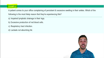
Introduction to the Lymphatic System Example 2
5263
views
99
rank
VideoThumbView.guidedCourse
3:33
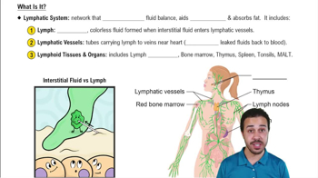
What Is It?
6915
views
182
rank
VideoThumbView.guidedCourse
3:57
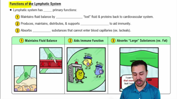
Functions of the Lymphatic System
5883
views
137
rank
이 집합의 용어 (31)
하이드의 정의
Main function of the lymphatic system
To return excess tissue fluid to the bloodstream and provide immune defense through lymphocytes and lymphoid organs.
Major lymph node areas in the body
Cervical, axillary, and inguinal lymph nodes.
General anatomy of a lymph node
Afferent vessels enter at the convex surface; efferent vessels exit at the concave surface (hilum).
Names and locations of tonsils
Palatine tonsils (oral cavity), pharyngeal tonsils/adenoids (nasopharynx), lingual tonsils (base of tongue), tubal tonsils (around auditory tube).
Location of Peyer’s patches
Isolated lymphatic nodules found in the small intestine.
Sign of active infection in a lymph node
Enlarged germinal centers in lymph follicles of the cortex, indicating dividing B cells.
Purpose and location of the thymus
Site where T lymphocytes develop immunocompetence; located mainly in the thorax, largest in children, atrophies after adolescence.
Fate of fetal thymus lymphocytes reactive to self-antigens
They are destroyed during selection to prevent autoimmune disease.
Thoracic duct origin and drainage
Begins at the cisterna chyli; drains lymph from left side of body and lower body into the junction of internal jugular and subclavian veins.
Right lymphatic duct drainage
Drains lymph from right upper torso into the junction of the right internal jugular and subclavian veins.
Function and location of the spleen
Site of RBC destruction and immune response; located in the left hypochondrium lateral to the stomach.
Innate immune system first line of defense examples
Intact skin (acid mantle, keratin), mucous membranes, nasal hairs, cilia, gastric juice, tears, saliva, urine.
Innate immune system second line of defense examples
Phagocytes, natural killer cells, inflammation, antimicrobial proteins, interferons, and fever.
Difference between humoral and cellular immunity
Humoral immunity involves B cells and antibody secretion; cellular immunity involves T cells targeting infected or abnormal cells.
Role of cytotoxic T lymphocytes
Attack virus-infected, cancer, or mismatched cells using MHC proteins; stimulated by helper T cells.
Types of immunity: passive natural
Antibodies received naturally, e.g., fetus receiving antibodies from mother.
Types of immunity: active artificial
Immune system produces antibodies after vaccination with weakened or dead pathogens.
Primary vs secondary immune response
Primary response has a 3-6 day lag on first antigen exposure; secondary response is faster and stronger due to immunological memory.
Mechanisms of antibody-antigen reaction
Neutralization, agglutination, precipitation, complement fixation, and cell lysis.
Functions of antibody classes IgM, IgA, IgD, IgG, IgE
IgM: first antibody in primary response; IgA: in secretions; IgD: B cell receptor; IgG: most abundant in plasma; IgE: triggers allergic reactions.
Effect of AIDS on immune system
Destruction of helper T lymphocytes, impairing immune response.
Definition and examples of autoimmune diseases
Immune system attacks self; examples include rheumatoid arthritis, lupus, multiple sclerosis, and Type 1 diabetes.
Cause of hypersensitive (allergic) reactions
IgE antibodies bind mast cells and basophils, triggering histamine release and inflammation.
Location of olfactory epithelium
Roof of the nasal cavity.
Cartilages of the larynx: paired and unpaired
Arytenoid cartilages are paired; epiglottis is elastic cartilage preventing food entry.
Largest airway with hyaline cartilage rings
The trachea.
Difference between right and left primary bronchi
Right bronchus is shorter, wider, and more vertical, making it more prone to lodging foreign objects.
Muscles responsible for inspiration
External intercostal muscles and diaphragm.
Relationship between PCO2, pH, and blood acidity
As PCO2 increases, pH decreases (more acidic); as PCO2 decreases, pH increases (more basic).
Definition and typical volume of tidal volume (TV)
Amount of air inhaled or exhaled during normal breathing; about 500 mL.
Function of surfactant in lungs
Produced by type II alveolar cells; reduces alveolar surface tension to prevent collapse.
 뒤로뒤로
 3:32