 뒤로
Anatomy & Physiology: Joints
 04:06

이 집합의 용어 (42)
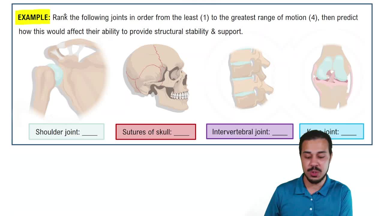
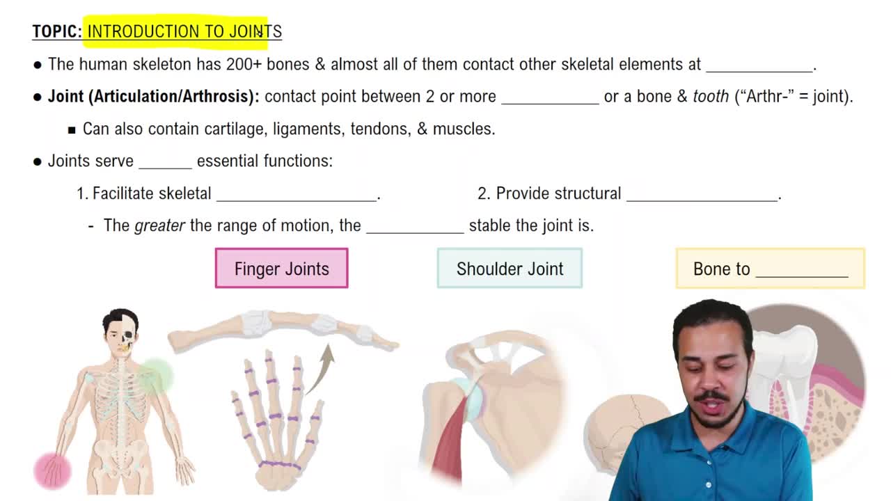
Joints provide skeleton mobility and hold the skeleton together.
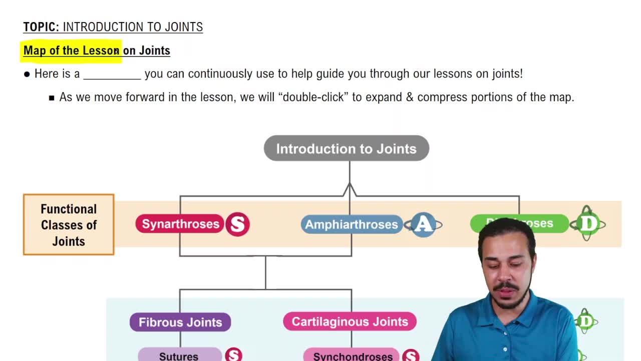
Synarthroses (immovable), Amphiarthroses (slightly movable), and Diarthroses (freely movable).
Synarthroses: immovable joints.
Amphiarthroses: slightly movable joints.
Diarthroses: freely movable joints.
Fibrous, Cartilaginous, and Synovial joints, based on material binding bones and presence of joint cavity.
Fibrous joints are bones joined by dense fibrous connective tissue with no joint cavity. Types: Sutures, Syndesmoses, and Gomphoses.
Rigid, interlocking joints with short connective tissue fibers, allowing growth in youth; ossify into synostoses in middle age.
Bones connected by ligaments; can be immovable or slightly movable. Examples include distal tibiofibular joint and interosseous membrane between radius and ulna.
Peg-in-socket fibrous joints of teeth in alveolar sockets, connected by the periodontal ligament.
Bones united by cartilage with no joint cavity. Types: Synchondroses (hyaline cartilage, immovable) and Symphyses (fibrocartilage, slightly movable).
Joints where bones are united by a bar or plate of hyaline cartilage; all are immovable (synarthrotic).
Joints where hyaline cartilage covers articulating surfaces fused to a pad of fibrocartilage; strong, flexible, and slightly movable (amphiarthrotic).
Freely movable (diarthrotic) joints with a joint cavity filled with synovial fluid; include most limb joints.
Articular cartilage, joint cavity, articular capsule (fibrous capsule and synovial membrane), synovial fluid, reinforcing ligaments, and rich nerve and blood supply.
Viscous filtrate of plasma and hyaluronic acid; lubricates and nourishes articular cartilage.
Bursae are fibrous sacs lined with synovial membrane containing fluid to reduce friction.
Tendon sheaths are elongated bursae that wrap around tendons.
Shape of articular surfaces, ligament number and location, and most importantly, muscle tone which keeps tendons taut.
Origin: attachment to immovable bone.
Insertion: attachment to movable bone.
Muscle contraction moves insertion toward origin.
One flat bone surface slips over another; occurs in intercarpal, intertarsal joints, and between vertebral articular processes.
Flexion: decreases joint angle.
Extension: increases joint angle.
Hyperextension: extension beyond normal range.
Abduction: movement away from midline.
Adduction: movement toward midline.
Circumduction: limb moves in a cone shape.
Turning of a bone around its long axis; occurs in joints like between C1 and C2 vertebrae, humerus, and femur.
Supination: palm faces forward.
Pronation: palm faces backward.
Dorsiflexion: upward foot movement.
Plantar flexion: downward foot movement.
Inversion: sole turns medially.
Eversion: sole turns laterally.
Elevation: lifting a body part superiorly.
Depression: moving a body part inferiorly.
Movement in the saddle joint allowing the thumb to touch the tips of other fingers.
Plane, hinge, pivot, condyloid, saddle, and ball-and-socket joints.
Nonaxial gliding movements; examples include intercarpal and intertarsal joints.
Uniaxial flexion and extension; examples include elbow and knee joints.
Uniaxial rotation; examples include the joint between C1 and C2 vertebrae and proximal radioulnar joint.
Biaxial angular movements like flexion, extension, abduction, and adduction; examples include metacarpophalangeal joints.
Biaxial joints with concave and convex articular surfaces; allow movements similar to condyloid joints but with greater freedom, e.g., carpometacarpal joint of the thumb.
Multiaxial movements including flexion, extension, abduction, adduction, and rotation; examples are shoulder and hip joints.
Stretching or tearing of ligaments; partial tears repair slowly, complete ruptures require surgical repair.
Fragments may cause joint locking; cartilage rarely repairs itself and is treated with arthroscopic surgery.
Bones forced out of alignment, often with sprains and inflammation, requiring joint immobilization.
Inflammation of bursae caused by blow or friction; treated with rest, ice, and anti-inflammatory drugs if severe.
Inflammation of tendon sheaths due to overuse; symptoms and treatment similar to bursitis.
Inflammatory or degenerative joint diseases causing pain, stiffness, and swelling.
Degenerative 'wear-and-tear' arthritis common in older adults, especially women; cartilage is destroyed faster than replaced.
Chronic autoimmune disease causing joint inflammation, pain, swelling, and eventual joint deformity.
Deposition of uric acid crystals causing inflammation, often affecting the base of the great toe; more common in men.