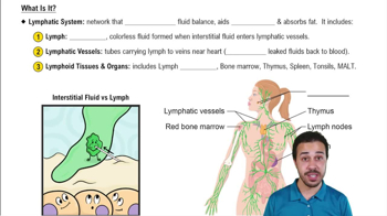
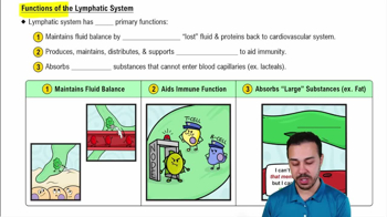
Regulation of interstitial fluid volume, absorption of dietary fats via lacteals, and immune functions by filtering pathogens and housing leukocytes.
What is lymph?
Fluid that exits extracellular space (interstitial fluid) and enters lymphatic vessels for transport back to the cardiovascular system.
Difference between lymphatic and blood capillaries
Lymphatic capillaries are blind-ended and one-way, allowing fluid and cells to enter but not exit; blood capillaries are two-way, moving blood toward and away from tissues.
Role of valves in lymphatic vessels
Prevent lymph from flowing backward, aiding transport against gravity, often assisted by muscle contractions.
Cisterna chyli and thoracic duct function
Cisterna chyli collects lymph from lumbar and intestinal trunks; thoracic duct drains lymph from lower body and left upper body into left subclavian vein.
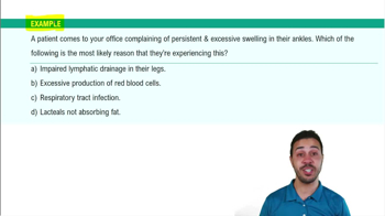
Lymphedema cause and effect
Caused by lymphatic vessel removal or blockage, leading to accumulation of interstitial fluid and swelling in affected body parts.
Composition of lymphoid tissue
Loose connective reticular tissue containing reticular fibers, macrophages, B and T lymphocytes, dendritic cells, and reticular cells.
Found in oral and nasal cavities, gastrointestinal tract (including Peyer's patches), respiratory passages, and genitourinary tract.
Function of lymph nodes
Filter pathogens from lymph, house leukocytes, and prevent spread of infection by trapping and destroying pathogens.
Spleen's two histological regions and functions
Red pulp destroys old erythrocytes; white pulp filters pathogens from blood and contains leukocytes.
Primary function of the thymus
Secretes hormones to generate functional T cells and ensures immunocompetence by screening T cells.
Three lines of defense in immunity
First line: surface barriers; second line: innate immunity (cells and proteins); third line: adaptive immunity (specific cells and antibodies).
Innate vs adaptive immunity
Innate immunity responds quickly and nonspecifically; adaptive immunity is slower, specific, and has immunological memory.
Surface barriers in first line of defense
Skin with keratin and acidic sebum; mucous membranes producing mucus and acid to trap and kill pathogens.
Phagocytic cells of innate immunity
Macrophages, neutrophils, and eosinophils that ingest and destroy pathogens and debris.
Role of natural killer (NK) cells
Nonphagocytic innate immune cells that recognize and destroy cancerous and virus-infected cells.
Complement system activation pathways
Classical (antibody-bound antigen), lectin (lectin binding carbohydrates), and alternative (direct pathogen recognition) pathways.
Main effects of activated complement proteins
Cell lysis via membrane attack complex, enhanced inflammation, virus neutralization, opsonization, and clearance of immune complexes.
Cardinal signs of inflammation
Redness, heat, swelling (edema), and pain caused by vasodilation, increased capillary permeability, and sensory neuron activation.
Phagocyte response stages in inflammation
Activation of local macrophages, neutrophil migration and diapedesis, monocyte recruitment and differentiation, leading to pathogen clearance.
Fever mechanism
Pyrogens reset hypothalamic thermostat to higher temperature, inducing shivering and heat production; fever breaks when reset to normal.
Cell-mediated immunity key cells
Helper T (TH) cells (CD4) and cytotoxic T (TC) cells (CD8) that respond to intracellular pathogens and abnormal cells.
Major histocompatibility complex (MHC) classes
Class I MHC on all nucleated cells present endogenous antigens to TC cells; Class II MHC on antigen-presenting cells present exogenous antigens to TH cells.
T cell activation requirements
Binding of T cell receptor to MHC-antigen complex and co-stimulation for full activation and clonal expansion.
Functions of helper T cells
Secrete cytokines to activate and enhance immune responses, including activation of TC cells and B cells.
Functions of cytotoxic T cells
Kill infected or abnormal cells by releasing perforin and enzymes inducing apoptosis.
 뒤로
 3:32