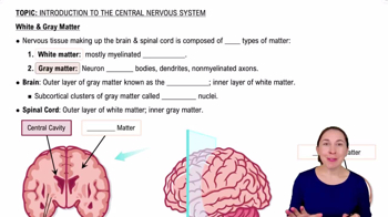
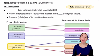
Central Nervous System (CNS) vs Peripheral Nervous System (PNS)
CNS includes brain and spinal cord, acting as integration and command center. PNS includes cranial and spinal nerves, serving as communication lines linking body parts to CNS.
Neuron Anatomy: Dendrites
Dendrites receive signals from other neurons and serve as the input region of the neuron.
Neuron Anatomy: Axon Hillock
The axon hillock is the trigger zone where graded potentials are summed and converted into an action potential if threshold is reached.
Functions of Astrocytes in CNS
Astrocytes support neurons, form the blood-brain barrier, and mop up excess ions and neurotransmitters.
Role of Oligodendrocytes vs Schwann Cells
Oligodendrocytes form myelin sheath in CNS; Schwann cells form myelin sheath in PNS.
Resting Membrane Potential
Established at about -70 mV mainly by the sodium-potassium pump pumping 3 Na+ out and 2 K+ in, plus K+ leak channels.
Action Potential Threshold
Threshold is around -55 mV at the axon hillock; if reached, an all-or-none action potential fires.
Voltage-Gated Sodium Channel States
Three states: Resting (closed), Activated (open) allowing Na+ influx, and Inactivated (blocked) during repolarization.
Cleared by enzymatic degradation, reuptake into presynaptic neuron or astrocytes, and diffusion away from synaptic cleft.
EPSP vs IPSP
EPSP is a graded depolarization pushing membrane potential closer to threshold; IPSP is a graded hyperpolarization pushing it further away.
Spatial vs Temporal Summation
Spatial summation: multiple presynaptic neurons fire simultaneously; Temporal summation: one neuron fires rapidly in succession.
Absolute vs Relative Refractory Period
Absolute: no action potential possible due to Na+ channel inactivation. Relative: action potential possible but requires stronger stimulus due to hyperpolarization.
Receptive Field Size and Sensitivity
Small receptive fields (e.g., fingertips) allow fine discrimination and high sensitivity; large fields (e.g., back) have low sensitivity and poor localization.
Tonic vs Phasic Receptors
Tonic receptors are always active and slow-adapting; Phasic receptors are fast-adapting and respond quickly then stop.
Five Receptor Types by Modality
Mechanoreceptors, Chemoreceptors, Thermoreceptors, Photoreceptors, and Nociceptors detect mechanical, chemical, temperature, light, and pain stimuli respectively.
High cGMP keeps Na+ channels open (dark current), depolarizing rods (~-40 mV) and causing continuous glutamate release inhibiting bipolar cells.
Phototransduction in the Light
Light bleaches rhodopsin → activates transducin → PDE breaks down cGMP → Na+ channels close → rod hyperpolarizes (~-70 mV) → glutamate release stops → bipolar cells activated.
Rhodopsin Cycle Steps
1. Bleaching/dissociation; 2. Transport of all-trans retinal to pigment epithelium; 3. Enzymatic isomerization to 11-cis retinal; 4. Recombination with opsin.
Why Dark Adaptation Takes Time
Rhodopsin must be regenerated via the rhodopsin cycle, which takes 20-30 minutes to restore night vision after bright light exposure.
Endolymph vs Perilymph Composition
Endolymph is high in K+ and low in Na+ (inside scala media); Perilymph is low in K+ and high in Na+ (scala vestibuli and tympani).
 뒤로
 0:50